Premolar
4,888 words · 16 min read
Quick Definition
A transitional tooth located between the canine and molar, featuring two cusps for crushing food. Adults have eight premolars, also called bicuspids.
Premolar: Your Essential Guide to These Powerhouse Teeth
Welcome to SmilePedia.net, your definitive resource for understanding every aspect of your oral health. Today, we're diving deep into the fascinating world of premolars, often known as bicuspids. These crucial teeth play a pivotal role in your ability to chew, speak, and maintain overall dental health. If you've ever wondered "what is a premolar?" or "why are premolar dental treatments important?", you're in the right place for a comprehensive answer.
Premolars are transitional teeth situated in the mid-mouth, perfectly positioned between your pointed canine teeth and your broader molar teeth. Their unique design, typically featuring two cusps (hence "bicuspid"), makes them highly effective at crushing and grinding food, preparing it for digestion. Every adult typically possesses eight premolars—two in each quadrant of the mouth, making them common and vital components of a healthy adult dentition. Understanding their function and common issues is key to preventing problems and preserving your smile for a lifetime.
Key Takeaways:
- Definition: Premolars (bicuspids) are transitional teeth located between canines and molars, primarily for crushing food.
- Number: Adults typically have eight premolars—two in each quadrant of the mouth.
- Anatomy: Characterized by two cusps (points) on their chewing surface.
- Function: Essential for effective mastication (chewing) and maintaining proper bite alignment.
- Dental Health: Prone to issues like cavities and fractures, requiring diligent oral hygiene and regular dental check-ups.
- Replacement: In children, premolars replace the primary (baby) molars as part of the permanent dentition.
What Are Premolars? Unpacking the Bicuspid
At the core of your chewing mechanism are your premolars, unique in their position and purpose. These teeth represent a transition zone, bridging the gap between the tearing action of your canines and the heavy grinding power of your molars. The term "bicuspid" provides a direct clue to their anatomy: "bi-" meaning two, and "-cuspid" referring to the pointed projections or eminences on the chewing surface of a tooth. Thus, most premolars typically feature two prominent cusps on their occlusal (chewing) surface – one on the buccal (cheek) side and one on the lingual (tongue) side.
In the complete adult dentition, you will find a total of eight premolars. There are two premolars in each of the four quadrants of your mouth:
- First Premolar: Closer to the front of the mouth, adjacent to the canine.
- Second Premolar: Located behind the first premolar, closer to the molars.
These teeth are classified as posterior teeth, alongside molars, meaning they are situated towards the back of the mouth. Their primary function is to crush and pulverize food, breaking it down into smaller, more manageable pieces before it reaches the powerful molars for final grinding. This intermediate crushing step is crucial for efficient digestion and plays a significant role in the overall biomechanics of your bite.
The Anatomy of a Premolar
Understanding the structure of a premolar helps to appreciate its function and vulnerability. Like all teeth, a premolar consists of a visible crown and an anchored root (or roots) embedded in the jawbone.
- Crown: This is the part you see in your mouth. The crown is covered by the hardest substance in the human body, enamel, which protects the softer inner layers.
- Occlusal Surface: The chewing surface of the premolar is characterized by its distinct cusps and grooves. Most premolars have two cusps (buccal and lingual), but some lower second premolars can have three (one buccal and two lingual). These cusps and the deep grooves (fissures) between them are critical for crushing food but also potential areas for plaque accumulation and cavities.
- Buccal Surface: The outer surface, facing the cheek.
- Lingual Surface: The inner surface, facing the tongue.
- Mesial and Distal Surfaces: The side surfaces that touch adjacent teeth.
- Dentin: Beneath the enamel lies dentin, a yellowish, bone-like tissue that makes up the bulk of the tooth. It contains microscopic tubules that can transmit sensations like hot and cold if the enamel is compromised.
- Pulp: At the core of the tooth is the pulp chamber and root canals, collectively known as the pulp. This soft tissue contains nerves, blood vessels, and connective tissue, providing vitality to the tooth.
- Root(s): The part of the tooth that anchors it into the jawbone. Most premolars have a single root, but the maxillary (upper) first premolar is unique among premolars in often having two roots. The roots are covered by cementum, a tissue that helps attach the tooth to the surrounding periodontal ligament and alveolar bone.
Types and Classifications of Premolars
While all premolars share the general function of crushing food, there are subtle yet important differences between them based on their position and arch (upper or lower jaw).
First Premolars (Bicuspids)
- Maxillary First Premolar (Upper Jaw): Often considered the most distinct of the premolars. It typically has two roots (a buccal and a palatal root), a feature more common in molars. Its cusps are usually sharper and more defined, with the buccal cusp being noticeably larger and longer than the lingual cusp. This tooth plays a significant role in providing space for orthodontic treatment when extractions are necessary.
- Mandibular First Premolar (Lower Jaw): Generally smaller than its maxillary counterpart and typically has only one root. The lingual cusp is often very small and non-functional, making it somewhat resemble a canine from an occlusal view, though it clearly has a buccal cusp and lingual cusp.
Second Premolars (Bicuspids)
- Maxillary Second Premolar (Upper Jaw): Typically has a single root, unlike the maxillary first premolar. Its two cusps are usually more equal in size and height, giving it a more rounded appearance. The grooves on the chewing surface are often less pronounced.
- Mandibular Second Premolar (Lower Jaw): This premolar is quite variable and can sometimes have three cusps (one buccal, two lingual), resembling a small molar. More commonly, it has two cusps that are more equal in size than the mandibular first premolar. It consistently has a single root.
Premolars in Primary vs. Permanent Dentition
It's important to note that children do not have premolars in their primary (baby) dentition. Instead, the spaces that will eventually house the permanent premolars are occupied by the primary molars. As children grow, their primary molars naturally exfoliate (fall out) and are replaced by the permanent premolars, typically between the ages of 10 and 12 years old. This transition is a crucial phase in the development of a strong, functional adult bite.
The Crucial Role of Premolars in Oral Health
Premolars are far more than just "filler" teeth; their presence and health are indispensable for a multitude of oral functions:
- Mastication (Chewing): This is their primary and most evident role. By crushing and tearing food into smaller particles, premolars significantly aid digestion. Without them, food would be less thoroughly broken down, potentially leading to digestive issues. They work in conjunction with the canines for initial tearing and the molars for final grinding.
- Maintaining Facial Structure: Along with other posterior teeth, premolars help support the vertical dimension of your bite and facial height. Their loss can lead to changes in facial appearance, such as a "collapsed" look or sagging in the lower face.
- Speech Articulation: While not as directly involved as front teeth, a full complement of teeth, including premolars, contributes to proper tongue and lip positioning, which is essential for clear speech.
- Arch Integrity and Alignment: Premolars act as important anchors within the dental arch. Their presence helps prevent adjacent teeth from shifting, drifting, or tilting, which can lead to bite problems, crooked teeth, and increased susceptibility to decay and gum disease. Losing a premolar can initiate a cascade of undesirable changes throughout your dentition.
Common Issues Affecting Premolars
Despite their robust design, premolars are susceptible to various dental problems that can compromise their function and your overall oral health.
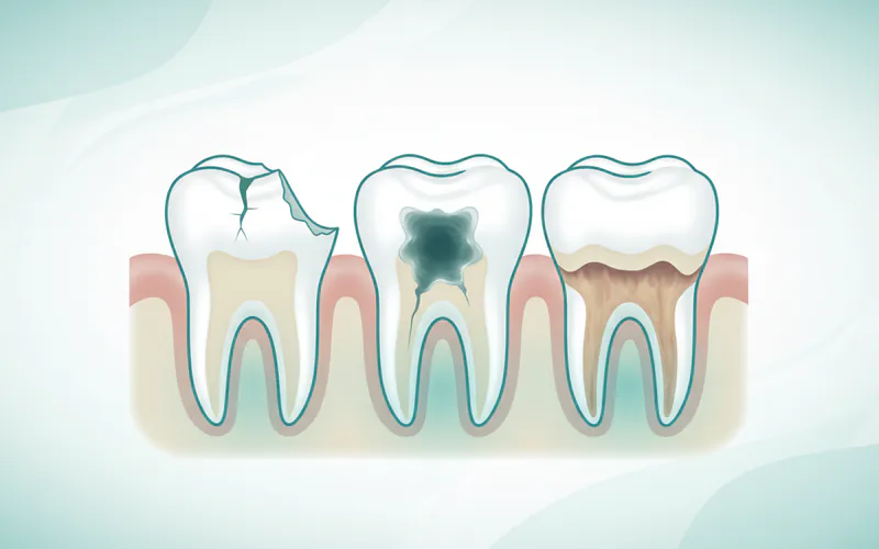
- Dental Caries (Cavities): Due to their chewing surfaces featuring cusps, pits, and fissures, premolars can easily trap food particles and plaque. If not cleaned effectively, this can lead to acid erosion and the formation of cavities. According to the CDC, over 25% of US adults aged 20-64 have untreated dental caries.
- Fractures and Cracks: The constant forces of chewing, especially on hard foods, can cause premolars to crack or fracture. Bruxism (teeth grinding or clenching) also puts immense pressure on these teeth, making them vulnerable. A cracked tooth can be incredibly painful and, if left untreated, can lead to infection.
- Impacted Premolars: While less common than impacted wisdom teeth, a premolar can sometimes fail to erupt properly and become trapped beneath the gum line or against an adjacent tooth. This can cause pain, infection, or damage to surrounding teeth.
- Periodontal Disease (Gum Disease): Like any tooth, premolars are susceptible to gum disease if plaque and tartar accumulate along the gum line. This can lead to inflammation, gum recession, bone loss, and ultimately, tooth mobility or loss.
- Sensitivity: Premolars can become sensitive to hot, cold, or sweet stimuli due to enamel erosion, gum recession exposing the root surface, or cracks.
- Tooth Loss: Severe decay, extensive trauma, or advanced periodontal disease can ultimately lead to the loss of a premolar, necessitating replacement options.
Signs and Symptoms to Watch For
Recognizing the early signs of a problem with your premolars can be crucial for timely and effective treatment. Be attentive to the following symptoms:
- Pain: This can manifest as a sharp pain when biting down, a throbbing ache that is constant, or a dull, persistent discomfort. Pain can indicate a cavity, a crack, an infection in the pulp, or gum inflammation.
- Sensitivity: Increased sensitivity to hot or cold foods and beverages, or even to sweet tastes, can signal enamel erosion, exposed dentin, or a developing cavity.
- Visible Changes: Look for dark spots, holes, or pits on the surface of the premolar, which are classic signs of a cavity. You might also notice a chip or crack.
- Swelling: Swelling in the gums around a premolar, or even facial swelling, can indicate an infection (abscess) in the tooth or surrounding tissues.
- Persistent Bad Breath or Bad Taste: An ongoing foul odor or taste in your mouth, despite good oral hygiene, could be a sign of a bacterial infection, often associated with decay or gum disease.
- Tenderness or Discomfort When Chewing: If biting down on a particular premolar causes pain or discomfort, it could be due to a cavity, a cracked tooth, or an inflamed pulp.
Diagnosis Process — What Your Dentist Does
If you experience any of the symptoms above, a visit to your dentist is essential. The diagnostic process for premolar issues is thorough and typically includes:
- Comprehensive Visual Examination: Your dentist will carefully inspect your premolars and surrounding gums, looking for any visible signs of decay, cracks, discoloration, swelling, or gum recession.
- Dental Probing: A small instrument called a periodontal probe is used to measure the depth of gum pockets around the teeth. Deeper pockets can indicate gum disease.
- Dental X-rays: This is a critical diagnostic tool.
- Bitewing X-rays: These are excellent for detecting cavities between teeth (interproximal decay) and assessing the health of the bone supporting your premolars.
- Periapical X-rays: These X-rays show the entire tooth, including the root and the surrounding bone, which is vital for diagnosing infections at the root tip or assessing root structure.
- Panoramic X-rays: Less specific for individual premolars but provides an overview of the entire jaw, useful for identifying impacted teeth or broader issues.
- Sensitivity Tests: If sensitivity or pain is a primary complaint, your dentist may use various tests (e.g., applying cold or heat, tapping on the tooth) to pinpoint the affected tooth and assess the vitality of the pulp.
- Palpation: Gentle pressure applied to the gums or jaw around the premolar can help identify areas of tenderness or swelling, indicating an infection.
- Transillumination: A focused light passed through the tooth can help reveal cracks that might not be visible otherwise.
Treatment Options for Premolar Issues
The treatment for a premolar issue depends entirely on the specific problem diagnosed. Here's an overview of common treatments, including their pros, cons, and typical cost ranges in the US.
1. Dental Fillings
- Purpose: To repair a premolar damaged by a cavity (dental caries).
- Procedure: The decayed portion of the tooth is removed, and the space is filled with a restorative material.
- Materials:
- Amalgam (silver) fillings: Durable and less expensive.
- Composite (tooth-colored) fillings: More aesthetically pleasing, bonded directly to the tooth.
- Gold or Porcelain: Less common for premolars due to cost and visibility.
- Pros: Preserves tooth structure, restores function, prevents further decay.
- Cons: Amalgam is visible; composite may not last as long as amalgam in high-stress areas; composite can stain.
- Cost Range (US): $50 - $250 per surface for amalgam; $100 - $450 per surface for composite.
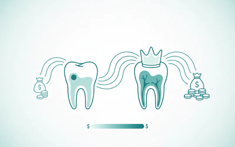
2. Dental Crowns (Caps)
- Purpose: To restore a premolar that has extensive decay, a large filling, a fracture, or has undergone root canal therapy.
- Procedure: The tooth is reshaped, impressions are taken, and a custom-made crown (often ceramic, porcelain-ffused-to-metal, or gold) is placed over the entire visible portion of the tooth.
- Pros: Strengthens and protects a weakened tooth, restores shape and function, aesthetically pleasing (especially ceramic).
- Cons: Requires reduction of healthy tooth structure, relatively expensive, can sometimes chip or fracture.
- Cost Range (US): $800 - $2,500 per crown, depending on material and location.
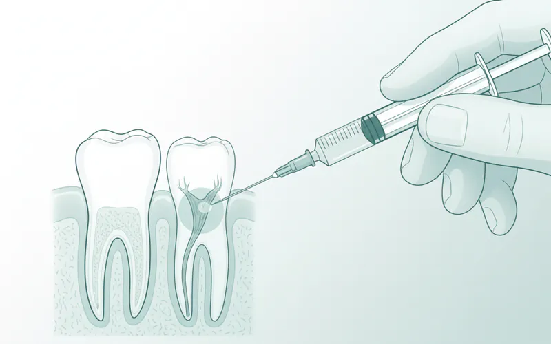
3. Root Canal Therapy (Endodontic Treatment)
- Purpose: To save a premolar whose pulp (nerve tissue) has become infected or inflamed due to deep decay, a crack, or trauma.
- Procedure: The infected pulp is removed from the pulp chamber and root canals, the canals are disinfected and shaped, and then filled with a biocompatible material. The tooth is typically then restored with a filling and often a crown.
- Pros: Saves the natural tooth, prevents extraction, resolves pain and infection.
- Cons: Can be a complex procedure, requires a follow-up restoration (often a crown) which adds to the cost, the tooth becomes brittle and requires protection.
- Cost Range (US): $700 - $1,500 for the root canal procedure itself, plus the cost of the crown.
4. Tooth Extraction
- Purpose: To remove a premolar that is severely damaged, untreatable by other means, or causing significant problems (e.g., severe infection, impaction, or for orthodontic space creation).
- Procedure: The tooth is carefully loosened from its socket and removed.
- Pros: Eliminates pain and infection immediately.
- Cons: Permanent loss of the tooth, can lead to shifting of adjacent teeth, bone loss in the jaw, and impacts chewing efficiency. Often requires replacement.
- Cost Range (US): $75 - $450 for a simple extraction; $150 - $650 for a surgical extraction.
5. Tooth Replacement (After Extraction)
- Purpose: To replace a missing premolar to restore function, aesthetics, and prevent shifting of other teeth.
- Options:
- Dental Implant: A titanium post surgically placed into the jawbone, serving as an artificial root, topped with a custom crown.
- Dental Bridge: A prosthetic tooth (pontic) anchored by crowns placed on the adjacent healthy teeth.
- Pros (Implants): Most natural feel, preserves jawbone, does not affect adjacent teeth.
- Cons (Implants): High cost, surgical procedure, longer treatment time.
- Pros (Bridges): Less invasive than implants, quicker to place.
- Cons (Bridges): Requires modifying adjacent healthy teeth, can put stress on anchor teeth, potential for decay under crowns, needs replacement over time.
- Cost Range (US):
- Dental Implant: $3,000 - $6,000+ per tooth (includes implant, abutment, and crown).
- Dental Bridge: $2,000 - $5,000 for a typical 3-unit bridge.
Step-by-Step: What to Expect During Treatment
While each treatment is unique, here’s a general idea of what to expect for common premolar procedures:
For a Dental Filling:
- Anesthesia: Local anesthetic will be administered to numb the area around the premolar, ensuring you feel no pain.
- Removal of Decay: Your dentist will use a drill to remove the decayed or damaged portion of the tooth.
- Preparation: The tooth is thoroughly cleaned and prepared for the filling material. For composite fillings, a bonding agent is applied.
- Filling Placement: The chosen filling material is applied to the tooth, shaped, and then hardened (with a special light for composite).
- Polishing: The filling is polished to ensure a smooth surface and proper bite.
For a Dental Crown:
- First Appointment (Preparation):
- Anesthesia: The area is numbed.
- Tooth Reshaping: The premolar is carefully reshaped to create space for the crown.
- Impressions: Molds of your teeth are taken to send to a dental lab for custom crown fabrication.
- Temporary Crown: A temporary crown is placed to protect the prepared tooth while your permanent crown is being made.
- Second Appointment (Placement):
- Temporary Removal: The temporary crown is removed.
- Permanent Crown Placement: Your dentist will check the fit, color, and bite of the new permanent crown.
- Cementation: Once satisfactory, the crown is permanently cemented onto your premolar.
For Root Canal Therapy:
- Anesthesia: Local anesthetic is given to numb the tooth and surrounding area.
- Isolation: A rubber dam is placed around the tooth to keep it dry and free of saliva during the procedure.
- Access Hole: A small opening is made in the crown of the premolar to access the pulp chamber and root canals.
- Pulp Removal: The infected or inflamed pulp tissue is carefully removed using specialized instruments.
- Cleaning and Shaping: The root canals are meticulously cleaned, disinfected, and shaped to prepare them for filling.
- Filling the Canals: The cleaned canals are filled with a biocompatible, rubber-like material called gutta-percha.
- Temporary Filling: A temporary filling is placed in the access hole.
- Final Restoration: A follow-up appointment is typically needed to place a permanent filling and, in most cases, a dental crown to protect the treated tooth.
Recovery Timeline and Aftercare
Recovery varies greatly depending on the treatment received:
- Fillings: Minimal recovery. You can usually eat normally once the numbness wears off (a few hours). Mild sensitivity for a few days is normal.
- Crowns: After the first appointment, you might experience mild sensitivity while wearing the temporary crown. After the permanent crown is placed, your tooth might be slightly sensitive for a few days as it adjusts. Full recovery is usually within a week.
- Root Canal Therapy: You might experience some tenderness or mild pain for a few days, which can be managed with over-the-counter pain relievers. Avoid chewing on the treated tooth until the final restoration (crown) is placed.
- Extraction: Expect some bleeding for the first day, and swelling and discomfort for 2-3 days. A soft diet is recommended for a few days. Avoid strenuous activity and smoking. Full healing of the socket takes several weeks.
General Aftercare Tips:
- Pain Management: Use over-the-counter pain relievers (ibuprofen, acetaminophen) as directed. Your dentist might prescribe stronger medication for more complex procedures.
- Soft Diet: Stick to soft foods for a few days, especially after extractions or root canals, to avoid irritating the treated area.
- Oral Hygiene: Continue brushing and flossing gently, avoiding the immediate treated area initially. Follow your dentist's specific instructions.
- Avoid Smoking and Alcohol: These can hinder healing.
- Follow-up Appointments: Attend all scheduled follow-up visits to ensure proper healing and address any concerns.
Prevention Strategies
Preventing premolar issues is always better (and less costly) than treating them. A consistent preventive routine can keep your premolars healthy for life:
- Excellent Oral Hygiene:
- Brush your teeth twice a day for two minutes each time, using a fluoride toothpaste. Pay special attention to the chewing surfaces of your premolars.
- Floss daily to remove plaque and food particles from between your teeth and under the gum line, areas where your toothbrush can't reach.
- Regular Dental Check-ups and Cleanings: Visit your dentist every six months for professional cleanings and examinations. These appointments allow your dentist to detect and address issues like cavities or gum disease in their early stages, often before they cause pain.
- Balanced Diet: Limit sugary and acidic foods and drinks, as these contribute to enamel erosion and cavity formation. Opt for a diet rich in fruits, vegetables, and lean proteins.
- Fluoride: In addition to fluoride toothpaste, consider using a fluoride mouthwash if recommended by your dentist. Professional fluoride treatments can also strengthen enamel.
- Dental Sealants: For children and sometimes adults with deep grooves (fissures) on their premolars, dental sealants can be applied. These are thin, protective plastic coatings painted onto the chewing surfaces to prevent food and bacteria from settling in the grooves and causing cavities.
- Mouthguards: If you grind or clench your teeth (bruxism) at night, a nightguard can protect your premolars from excessive wear and potential fractures. For athletes, a custom-fitted sports mouthguard is essential to protect teeth from trauma during contact sports.
Cost Ranges in the US (With/Without Insurance)
Understanding the financial aspect of premolar treatments is important. Costs can vary significantly based on your location, the dentist's fees, the complexity of the procedure, and the materials used.
- Dental Fillings:
- Amalgam: $50 - $250 per surface
- Composite: $100 - $450 per surface
- Dental Crowns: $800 - $2,500 per crown
- Root Canal Therapy: $700 - $1,500 (plus the cost of a crown)
- Simple Extraction: $75 - $450
- Surgical Extraction: $150 - $650
- Dental Implant (Single Tooth): $3,000 - $6,000+
- Dental Bridge (3-unit): $2,000 - $5,000
Insurance Coverage: Most dental insurance plans provide some level of coverage for preventive and restorative procedures.
- Preventive Care (cleanings, exams, X-rays): Often covered at 80-100%.
- Basic Restorative Care (fillings, simple extractions): Typically covered at 50-80%.
- Major Restorative Care (crowns, root canals, bridges): Usually covered at 30-50%.
- Implants: Coverage varies widely; some plans offer minimal or no coverage, while others cover 20-50%.
It's crucial to check with your specific dental insurance provider to understand your benefits, deductibles, and annual maximums. Your dentist's office can often help you navigate insurance claims and provide estimated out-of-pocket costs.
Comparison Table: Common Premolar Treatments
| Treatment | Purpose | Durability | Cost (US Range, without insurance) | Pros | Cons |
|---|---|---|---|---|---|
| Dental Filling | Repair minor to moderate decay | Amalgam: 10-15+ years; Composite: 5-10 years | $50 - $450 | Preserves tooth, relatively quick, cost-effective | Amalgam is visible; Composite may not be as durable as amalgam in high-stress areas |
| Dental Crown | Restore extensively damaged/weakened tooth | 10-15+ years | $800 - $2,500 | Strengthens and protects, restores full function, good aesthetics | Requires tooth reduction, higher cost, requires multiple visits |
| Root Canal | Save tooth with infected/inflamed pulp | Many years, with proper crown | $700 - $1,500 (plus crown) | Saves natural tooth, prevents extraction, resolves infection/pain | Can be complex, tooth becomes brittle (requires crown), higher overall cost with crown |
| Extraction | Remove severely damaged/untreatable tooth | Permanent removal | $75 - $650 | Immediate relief from severe pain/infection | Permanent tooth loss, can lead to shifting/bone loss, requires replacement for optimal function |
| Dental Implant | Replace single missing tooth (after extraction) | 20+ years, often lifetime | $3,000 - $6,000+ | Most natural, preserves bone, doesn't affect adjacent teeth | High cost, surgical procedure, longer treatment time, not suitable for all patients |
| Dental Bridge | Replace 1-2 missing teeth (after extraction) | 5-15 years | $2,000 - $5,000 | Quicker than implant, less invasive than surgery | Requires modification of healthy adjacent teeth, can cause stress on anchor teeth, needs eventual replacement |
For Parents / Pediatric Considerations
While children don't have permanent premolars until adolescence, the care of their primary (baby) teeth is critical to the healthy development and eruption of their adult premolars.
- Primary Molars as Precursors: The first and second primary molars are the predecessors to the permanent first and second premolars, respectively. Maintaining the health of these baby molars prevents premature loss, which can lead to space loss and misalignment issues for the erupting permanent premolars.
- Eruption Timeline: Permanent premolars typically erupt between ages 10 and 12, replacing the primary molars. Parents should monitor this process for any signs of delayed eruption or crowding.
- Sealants for New Premolars: Once permanent premolars erupt, applying dental sealants to their deep grooves and fissures can provide an excellent defense against cavities, just as they do for molars. This is a simple, painless procedure that creates a protective barrier.
- Orthodontic Considerations: Premolars are sometimes strategically extracted during orthodontic treatment (braces) to create necessary space for overcrowded teeth or to correct severe bite issues. This decision is made by an orthodontist after careful assessment.
Frequently Asked Questions
How much does premolar treatment cost?
Costs vary widely depending on the type of treatment needed. A simple filling might cost $50-$450, while a root canal and crown can range from $1,500-$4,000. Replacing a missing premolar with a dental implant typically costs $3,000-$6,000+. Dental insurance usually covers a portion of these costs, often 50-80% for basic restorative care and 30-50% for major procedures, after deductibles are met.
Is it painful to have work done on a premolar?
Thanks to local anesthesia, you should not feel any pain during most premolar treatments (like fillings, crowns, or root canals). You might feel pressure or vibration, but no sharp pain. After the anesthesia wears off, some discomfort or sensitivity is common, which can usually be managed with over-the-counter pain relievers. Your dentist will provide specific post-treatment care instructions.
How long does a premolar filling or crown last?
The lifespan varies by material and oral hygiene. Amalgam (silver) fillings can last 10-15 years or more, while composite (tooth-colored) fillings typically last 5-10 years. Dental crowns are quite durable, often lasting 10-15 years, and sometimes even longer, with good care. Regular check-ups and good oral hygiene are key to maximizing their longevity.
Can I live without a premolar?
While you can technically "live" without a premolar, its loss can lead to several problems. The adjacent teeth may shift, drift, or tilt into the empty space, disrupting your bite and potentially causing alignment issues. The opposing tooth in the other jaw can also super-erupt (grow longer) without an opposing tooth to bite against. This can lead to chewing difficulties, gum problems, and temporomandibular joint (TMJ) issues. Replacing a missing premolar is generally recommended to maintain oral health and function.
What are the alternatives to extracting a damaged premolar?
If a premolar is damaged, extraction is usually a last resort. Alternatives often include:
- Dental Filling: For small to moderate cavities.
- Dental Crown: For larger cavities, fractures, or after a root canal to protect the tooth.
- Root Canal Therapy: To save a tooth with an infected or inflamed pulp. Your dentist will assess the extent of the damage to determine if the tooth can be saved.
Does dental insurance cover premolar treatments?
Most dental insurance plans provide some coverage for premolar treatments, but the extent of coverage depends on your specific plan. Preventive care (like exams and cleanings) is often fully covered. Basic restorative treatments (like fillings and simple extractions) typically have 50-80% coverage, while major procedures (like crowns, root canals, and bridges) usually fall into the 30-50% coverage category. Coverage for dental implants varies significantly. Always consult your insurance provider for details.
Why are premolars sometimes removed for orthodontics?
In some orthodontic cases, particularly when there is severe crowding or significant bite discrepancies, one or more premolars might be extracted. This creates necessary space within the dental arch, allowing the orthodontist to align the remaining teeth properly, reduce protrusion, and achieve a stable bite. The decision to extract premolars is made carefully by an orthodontist after a thorough analysis of the patient's specific needs.
When to See a Dentist
Your premolars are vital for your oral health, and prompt attention to any issues can prevent more serious problems.
See a dentist immediately (Emergency) if you experience:
- Severe, throbbing pain that doesn't subside.
- Significant swelling in your gums or face.
- A tooth that feels loose or has been knocked out.
- Trauma to your premolar from an accident.
Schedule an urgent appointment if you have:
- Persistent sensitivity to hot, cold, or sweets.
- A visible hole or dark spot on your premolar.
- A lost filling or crown.
- Pain when biting or chewing.
- Bleeding or tender gums around a premolar.
Regular routine care is essential:
- Visit your dentist every six months for comprehensive check-ups and professional cleanings. These routine visits are the best way to catch and address problems with your premolars (and all your teeth) before they become painful or severe.
Medical Disclaimer
This article is for informational purposes only and does not constitute medical advice. Always consult with a qualified dental professional for diagnosis and treatment. Do not delay seeking professional advice because of something you read on this website.