Beginning Oral Cancer Stages: Complete Guide
Key Takeaways
- Imagine discovering a small, seemingly harmless patch in your mouth that could silently be a precursor to a life-altering disease. Oral cancer, often overlooked in its initial phases, is a formidable health challenge. Did you know that early detection dramatically increases survival rates, with over
Beginning Oral Cancer Stages: Complete Guide
Imagine discovering a small, seemingly harmless patch in your mouth that could silently be a precursor to a life-altering disease. Oral cancer, often overlooked in its initial phases, is a formidable health challenge. Did you know that early detection dramatically increases survival rates, with over 80% of cases being treatable when caught in the beginning oral cancer stages? Unfortunately, many cases are diagnosed only after the disease has advanced, leading to more complex treatments and a lower prognosis.

This article, crafted by the experts at SmilePedia.net, is your definitive guide to understanding the crucial early signs, diagnosis, and preventative measures against oral cancer. We will delve into what oral cancer is, explore its various forms, uncover the primary causes, and equip you with the knowledge to recognize subtle symptoms. From detailing the comprehensive oral cancer screening process to demystifying treatment options and associated costs, our aim is to empower you with the information needed to protect your oral and overall health. Understanding these initial stages is not just about awareness; it's about enabling timely intervention that can truly save lives.
Key Takeaways:
- Early Detection is Crucial: Oral cancer survival rates exceed 80-90% when diagnosed in the beginning stages (Stage 0 or I), significantly dropping to around 50-60% for advanced stages.
- Common Early Signs: Look for persistent red (erythroplakia) or white (leukoplakia) patches, non-healing sores (lasting over 2 weeks), lumps, or unexplained numbness in the mouth.
- Oral Cancer Screenings: Regular screenings, ideally annually, are vital for early detection. The American Dental Association (ADA) recommends comprehensive oral exams as part of routine dental check-ups.
- Screening Costs: A typical visual and tactile oral cancer screening is often included in a routine dental exam, costing $50-$200 without insurance if performed separately. Adjunctive technologies may add $25-$75.
- Biopsy Confirms Diagnosis: If a suspicious lesion is found, a biopsy is the definitive diagnostic tool. An incisional biopsy typically costs between $300-$800 (without insurance) for the procedure and pathology.
- HPV Link: Human Papillomavirus (HPV) is a growing risk factor, especially for oropharyngeal cancers. Vaccination can offer protection.
- Lifestyle Matters: Avoiding tobacco (all forms) and excessive alcohol consumption are the most impactful preventive measures, reducing risk by up to 80%.
What Oral Cancer Is: An Overview
Oral cancer, often referred to as mouth cancer, is a type of head and neck cancer that can develop in any part of the oral cavity. This includes the lips, tongue, floor of the mouth, inner lining of the cheeks, gums, roof of the mouth (hard and soft palate), and the oropharynx (the part of the throat just behind the mouth, including the tonsils and base of the tongue). It typically originates from the squamous cells that line the moist surfaces inside the mouth and throat, hence the most common type is squamous cell carcinoma.
The disease begins when cells in the oral cavity start to grow uncontrollably, forming tumors or lesions. If left untreated, these cancerous cells can spread from their original location (local invasion) to nearby lymph nodes in the neck and, eventually, to other parts of the body (metastasis), making treatment much more challenging and reducing the chances of survival.
Prevalence and Impact: According to the Oral Cancer Foundation, close to 54,000 Americans are diagnosed with oral or oropharyngeal cancer each year. While it affects both men and women, it is historically more common in men, particularly those over 40. However, in recent years, there has been a concerning rise in diagnoses among younger individuals, largely attributed to the increasing prevalence of Human Papillomavirus (HPV) infection. The disease claims one life every hour of every day in the U.S., highlighting the critical need for awareness and early detection. The good news is that when identified in its beginning oral cancer stages, the survival rate can be as high as 85-90%, underscoring why understanding and recognizing early signs is paramount.
Types of Oral Cancer and Variations
While squamous cell carcinoma accounts for about 90% of all oral cancers, it's important to understand where and how these cancers can manifest, as location and appearance can sometimes hint at the type or aggressiveness.
Squamous Cell Carcinoma (SCC)
This is the most common form, originating from the flat, thin cells (squamous cells) that line the mouth and throat. SCC can appear as a persistent sore, a red or white patch, a lump, or an erosion that doesn't heal. It can develop on any oral surface, but is most frequently found on the tongue (especially the sides), the floor of the mouth, the soft palate, and the tonsils.
Other Less Common Types:
- Minor Salivary Gland Cancers: These can occur anywhere in the mouth where minor salivary glands are present, often on the hard or soft palate. Types include adenoid cystic carcinoma, mucoepidermoid carcinoma, and polymorphous adenocarcinoma.
- Lymphomas: These cancers arise in lymph tissue and can sometimes affect the tonsils or the base of the tongue.
- Melanoma: Though rare, melanoma can develop in the mouth, usually appearing as a dark, pigmented lesion, similar to skin melanoma.
- Sarcomas: These cancers arise in bone or soft tissue and are extremely rare in the oral cavity.
Precancerous Lesions (Dysplasia)
Recognizing precancerous changes is vital for intercepting oral cancer before it fully develops. These lesions indicate abnormal cell growth that isn't yet cancerous but has the potential to become malignant.
- Leukoplakia: These are white or grayish patches that cannot be rubbed off. They are typically found on the tongue, floor of the mouth, or inner cheek. While most leukoplakia patches are benign, about 5-20% can develop into SCC, especially if they are speckled (erythroleukoplakia) or have an irregular texture.
- Erythroplakia: These are red, velvety patches that are often flat or slightly depressed. Erythroplakia is much less common than leukoplakia but carries a significantly higher risk of malignancy, with up to 70-90% of these lesions being severely dysplastic or already cancerous. Their intense redness often signifies increased blood supply to abnormal cells.
- Erythroleukoplakia (Speckled Leukoplakia): This refers to lesions that are a mix of red and white. These are particularly concerning due to their high potential for malignancy, combining the risks of both leukoplakia and erythroplakia.
- Oral Submucous Fibrosis: A chronic, progressive, debilitating disease of the oral cavity characterized by inflammation and progressive fibrosis of the submucosal tissues. It is primarily associated with betel quid chewing and can lead to restricted mouth opening and is considered a highly precancerous condition.
Causes and Why It Happens
Oral cancer, like most cancers, results from damage to the DNA within cells, leading to uncontrolled growth. Several risk factors significantly increase the likelihood of these genetic mutations. Understanding these causes is crucial for prevention.
Primary Risk Factors:
- Tobacco Use: This is by far the leading cause, responsible for an estimated 80% of oral cancers. This includes smoking cigarettes, cigars, pipes, and using smokeless tobacco products (chewing tobacco, dip, snuff). The carcinogens in tobacco directly damage the cells in the mouth, leading to abnormal growth. The risk is dose-dependent and cumulative, increasing with the duration and amount of tobacco used.
- Alcohol Consumption: Heavy and chronic alcohol use is another major risk factor. Alcohol acts as an irritant to oral tissues and can also enhance the penetration of other carcinogens (like those in tobacco) into the cells. The combination of tobacco and alcohol creates a synergistic effect, meaning the risk is much higher than the sum of the individual risks. For individuals who both smoke and drink heavily, the risk of developing oral cancer can be up to 30 times higher than for non-users.
- Human Papillomavirus (HPV) Infection: Certain high-risk strains of HPV, particularly HPV-16, are a rapidly growing cause of oropharyngeal cancers (cancers of the tonsils and base of the tongue). Unlike tobacco- and alcohol-related cancers which often affect older individuals, HPV-related oral cancers are increasingly seen in younger, non-smoking, non-drinking adults. This virus is primarily transmitted through oral sexual contact.
- Excessive Sun Exposure: For cancers of the lip, particularly the lower lip, prolonged and unprotected exposure to ultraviolet (UV) radiation from the sun is a significant risk factor, similar to skin cancer.
- Age: The risk of oral cancer increases with age, with most diagnoses occurring in individuals over 40 years old.
- Weakened Immune System: People with compromised immune systems (e.g., organ transplant recipients, individuals with HIV/AIDS) are at a higher risk.
- Poor Nutrition: A diet consistently low in fruits and vegetables and high in processed foods may contribute to an increased risk.
- Genetic Predisposition: While less common, a family history of oral or other head and neck cancers can slightly increase an individual's risk.
- Chronic Irritation (less definitive): While historically thought to be a cause, chronic irritation from ill-fitting dentures, jagged teeth, or fillings is generally not considered a direct cause of oral cancer. However, such irritation can create persistent sores that should be monitored, as a non-healing ulcer is a significant symptom.
Pro Tip: If you use tobacco or consume alcohol regularly, discuss your risk factors with your dentist. They can provide resources for cessation and emphasize the importance of more frequent oral cancer screenings.
Signs and Symptoms: What to Look For in Beginning Oral Cancer Stages
Recognizing the subtle warning signs in the beginning oral cancer stages is paramount for successful treatment. Many early symptoms are painless or mimic less serious conditions, making vigilance crucial. The key is persistence – if any of these signs last longer than two weeks, it warrants immediate professional evaluation.
Visual Signs:
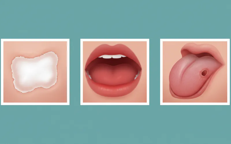
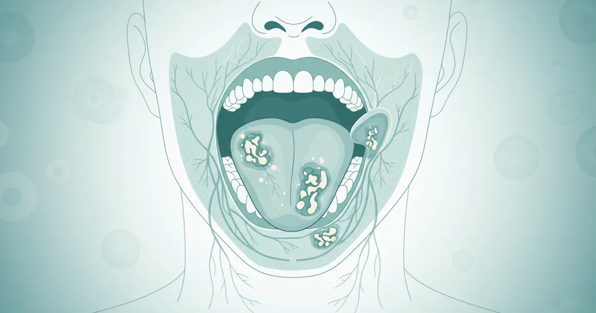
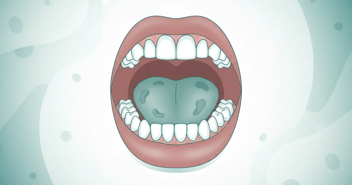
- White Patches (Leukoplakia): These are common, usually harmless, but some can be precancerous. They appear as flat, white, or grayish areas on the tongue, inner cheeks, gums, or floor of the mouth that cannot be scraped off. They may feel slightly thickened or rough.
- Red Patches (Erythroplakia): These are less common but far more concerning. They appear as bright red, velvety areas that can be flat or slightly raised. They often bleed easily and are highly indicative of dysplasia or early cancer.
- Mixed Red and White Patches (Erythroleukoplakia): These speckled patches combine the characteristics of both leukoplakia and erythroplakia and carry a very high risk of malignancy.
- Non-Healing Sore or Ulcer: This is one of the most classic signs. Any sore, lesion, or ulcer in the mouth or on the lips that does not heal within two weeks should be considered suspicious. It may be painless initially but can become painful as it progresses.
- Lump, Thickening, or Swelling: Feeling a palpable lump or an area of thickened tissue on the lip, gum, tongue, or elsewhere inside the mouth. These may not be painful.
- Unusual Bleeding: Any unexplained bleeding from the mouth or gums that is not due to trauma or gum disease.
Non-Visual and Sensory Symptoms:
- Persistent Sore Throat or Hoarseness: A chronic sore throat, a feeling that something is caught in your throat, or a persistent change in voice quality (hoarseness) could indicate cancer in the oropharynx or larynx.
- Difficulty Swallowing (Dysphagia) or Chewing: Pain or difficulty when swallowing food or liquids (dysphagia) or chewing, possibly due to a lesion obstructing the passage or causing discomfort.
- Difficulty Moving the Tongue or Jaw: Reduced mobility of the tongue or jaw, or pain when moving them. This can indicate involvement of deeper tissues.
- Numbness or Pain: Persistent numbness (paresthesia) or pain in any area of the mouth, face, or neck without an obvious cause. While early lesions may be painless, localized pain can develop.
- Persistent Earache: A persistent earache in one ear, especially when accompanied by jaw pain or difficulty swallowing, can be a referred pain symptom of advanced oral or oropharyngeal cancer.
- Loose Teeth or Dentures that No Longer Fit: If teeth become unexpectedly loose without a clear dental reason (like periodontal disease), or if dentures start to fit poorly and cause irritation, it could be a sign of cancer invading the jawbone or gums.
- Unexplained Weight Loss: Significant, unintentional weight loss can be a symptom of many types of cancer, including oral cancer, especially if eating is painful or difficult.
- Changes in Taste: A persistent alteration in taste sensation or a metallic taste in the mouth.
Pro Tip: Perform a self-examination monthly! Stand in front of a mirror with good lighting. Use your fingers to feel for lumps or abnormal textures on your lips, cheeks, gums, tongue (top, sides, and underneath), and the floor and roof of your mouth. Pull your tongue out and look at its sides and base. Look for any changes in color (redness, whiteness, dark spots) or any sores that haven't healed.
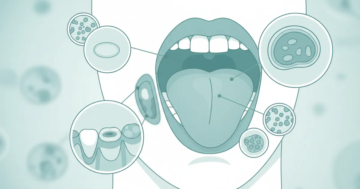
Oral Cancer Screening: The First Line of Defense
Regular oral cancer screening is the single most important step in detecting the disease in its beginning oral cancer stages. The American Dental Association (ADA) recommends that all adults receive a comprehensive oral examination, which includes an oral cancer screening, at least annually as part of their routine dental check-up.
What to Expect During a Screening:
A typical oral cancer screening is quick, painless, and integrated into your regular dental visit.
- Visual Examination: Your dentist will carefully inspect your entire oral cavity and surrounding areas.
- External Examination: They'll look at your face, neck, and lips for any asymmetry, swelling, lumps, or unusual skin changes.
- Internal Examination: Using a light and a mirror, they'll systematically examine the inside of your mouth: your tongue (top, sides, and underside), gums, inner cheeks, roof of your mouth, and the floor of your mouth. They'll look for any of the signs discussed previously – red or white patches, sores, lumps, or unusual textures.
- Tactile Examination (Palpation): Your dentist will use their gloved fingers to feel for any lumps, hardened areas, or tenderness in your mouth, under your jaw, and along your neck. They will often ask you to stick out your tongue and move it from side to side to check for mobility.
- Medical History Review: Your dentist will also review your medical history and discuss risk factors such as tobacco and alcohol use, sun exposure, and family history.
Adjunctive Screening Technologies:
While the visual and tactile exam remains the gold standard, several adjunctive technologies can help dentists identify suspicious areas that might be missed by the naked eye, or to better assess existing lesions. These are often used as supplemental tools, not replacements for a thorough clinical exam.
- Toluidine Blue Dye: This special blue dye is rinsed around the mouth or applied directly to suspicious areas. Abnormal cells absorb the dye more intensely, making precancerous or cancerous lesions stand out as darker blue spots. This can help define lesion margins.
- Light-Based Detection Systems (e.g., VELscope, Identafi, ViziLite): These devices use specific wavelengths of light to visualize changes in oral tissue that might not be visible under normal white light.
- Fluorescence Visualization (FV): Devices like VELscope emit a blue light. Healthy oral tissue naturally fluoresces (glows green), while abnormal tissue (due to changes in collagen and cellular metabolism) loses its fluorescence and appears dark.
- Reflectance Imaging: Other systems use different lights (e.g., white, green, violet) to highlight specific tissue characteristics, such as increased vascularity or changes in epithelial thickness, that can indicate abnormal cell activity.
- Brush Biopsy (OralCDx): This technique involves using a small brush to collect cells from a suspicious area. The cells are then sent to a lab for microscopic analysis. While less invasive than a traditional scalpel biopsy, it's often used for initial assessment of seemingly benign lesions and, if positive, still requires a scalpel biopsy for definitive diagnosis.
Pro Tip: Don't hesitate to ask your dentist about oral cancer screening during your regular check-up, especially if you have risk factors or notice any persistent changes in your mouth.
Diagnosis: Beyond the Screening
If your dentist identifies a suspicious area during a screening, the next crucial step is to obtain a definitive diagnosis. This usually involves a referral to an oral surgeon, oral pathologist, or an ENT (Ear, Nose, and Throat) specialist, who will perform a biopsy.
The Biopsy: Definitive Diagnosis
A biopsy is the removal of a small tissue sample from the suspicious area for microscopic examination by a pathologist. This is the only way to confirm the presence of cancer or precancerous cells.
- Incisional Biopsy: This is the most common type. A small wedge of tissue is surgically removed from the abnormal area under local anesthesia. The excised tissue includes a portion of both the suspicious lesion and some healthy surrounding tissue.
- Excisional Biopsy: If the suspicious lesion is small and easily accessible, the entire lesion may be removed during the biopsy. This can sometimes serve as both diagnosis and initial treatment.
- Punch Biopsy: A circular blade is used to remove a small core of tissue.
- Brush Biopsy (Cytology): As mentioned, this involves collecting surface cells with a brush. While useful for initial screening, if positive, it usually necessitates a scalpel biopsy for a more in-depth, definitive diagnosis, as it only samples superficial layers.
The tissue sample is then processed and examined under a microscope by an oral pathologist. They will determine if the cells are benign, precancerous (dysplastic), or cancerous, and if cancerous, what type and grade (how aggressive it appears).
Staging of Oral Cancer: Determining Extent
Once cancer is confirmed, additional tests are performed to determine the stage of the cancer. Staging describes the size of the tumor, whether it has spread to nearby lymph nodes, and if it has metastasized to distant parts of the body. The most common system is the TNM staging system:
- T (Tumor): Refers to the size and extent of the primary tumor.
- N (Nodes): Indicates whether the cancer has spread to nearby lymph nodes.
- M (Metastasis): Denotes whether the cancer has spread to distant organs.
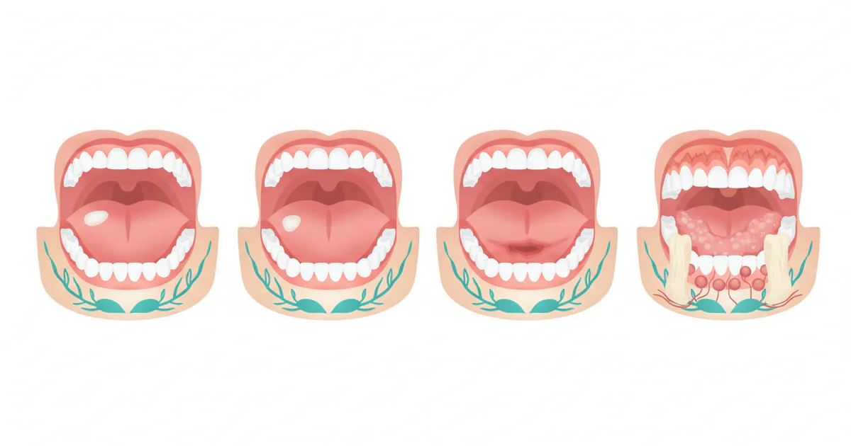
Beginning Oral Cancer Stages typically fall into:
- Stage 0 (Carcinoma in Situ): Abnormal cells are present only in the top layer of cells and have not invaded deeper tissues. This is highly treatable.
- Stage I: The tumor is small (2 cm or less) and has not spread to lymph nodes or distant sites. Early detection at these stages is why prognosis is so favorable. Imaging tests like CT scans, MRI, or PET scans are used to accurately assess the extent of the disease for staging purposes.
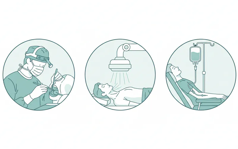
Treatment Options
The treatment plan for oral cancer depends heavily on the stage of the cancer, its location, the patient's overall health, and personal preferences. For beginning oral cancer stages (Stage 0, I, and some Stage II), treatment is generally less aggressive and highly successful.
1. Surgery
Surgery is often the primary treatment for early-stage oral cancers.
- Primary Tumor Excision: The tumor and a margin of healthy tissue around it (to ensure all cancer cells are removed) are surgically excised. This can be done using a scalpel, laser, or cryosurgery (freezing).
- Neck Dissection: If there's a risk of cancer spreading to the lymph nodes (even if not clinically evident), or if lymph nodes are already involved, lymph nodes in the neck may be removed. This is often done proactively to prevent spread or to remove existing spread.
- Reconstructive Surgery: For larger resections, reconstructive surgery (using tissue from other parts of the body) may be necessary to restore function (speech, swallowing) and appearance.
Pros: Often curative for early stages; immediate removal of cancerous tissue. Cons: Can be disfiguring, may require reconstructive surgery, potential for speech and swallowing difficulties.
2. Radiation Therapy
Radiation therapy uses high-energy rays (like X-rays or protons) to kill cancer cells.
- External Beam Radiation Therapy (EBRT): A machine outside the body directs radiation to the tumor and surrounding areas. This is usually given daily over several weeks.
- Brachytherapy: Radioactive seeds or wires are temporarily or permanently placed directly into or near the tumor. This delivers a high dose of radiation directly to the cancer while minimizing exposure to healthy tissues.
Pros: Can preserve organ function; non-invasive; effective for localized cancers. Cons: Side effects can include dry mouth (xerostomia), mucositis (sore mouth), taste changes, jaw stiffness (trismus), and long-term risk of osteoradionecrosis (bone death).
3. Chemotherapy
Chemotherapy uses drugs to kill cancer cells, either by stopping their growth or by destroying them directly.
- Adjuvant Chemotherapy: Given after surgery to kill any remaining cancer cells.
- Neoadjuvant Chemotherapy: Given before surgery or radiation to shrink the tumor.
- Concomitant Chemoradiation: Chemotherapy and radiation are given together, as chemotherapy can make cancer cells more sensitive to radiation.

Pros: Can treat cancer that has spread or is widespread. Cons: Significant systemic side effects (nausea, fatigue, hair loss, weakened immune system, mouth sores).
4. Targeted Therapy
These drugs specifically target unique features of cancer cells (like certain proteins or genetic mutations) that help them grow and spread, minimizing harm to healthy cells. For oral cancer, targeted therapies often focus on epidermal growth factor receptor (EGFR) inhibitors.
Pros: Fewer side effects than traditional chemotherapy; more precise action. Cons: Only effective for cancers with specific targets; can be very expensive.
5. Immunotherapy
Immunotherapy harnesses the body's own immune system to fight cancer. Drugs called checkpoint inhibitors block proteins that prevent immune cells from attacking cancer cells, essentially "releasing the brakes" on the immune response.
Pros: Can provide long-lasting responses in some patients; effective in certain advanced cases. Cons: Can cause immune-related side effects; not effective for all patients; very expensive.
Treatment Option Comparison for Early-Stage Oral Cancer
| Treatment Option | Primary Use for Early Stages | Pros | Cons |
|---|---|---|---|
| Surgery (Excision) | Most common for Stage 0, I, and some II primary tumors. | High success rate in removing localized cancer; immediate results. | Can be invasive; potential for cosmetic and functional changes; requires recovery time. |
| Radiation Therapy | Alternative to surgery for small tumors, or adjuvant after surgery. | Non-invasive (EBRT); can preserve organ function; effective for hard-to-reach areas. | Multiple sessions over weeks; short-term side effects (mucositis, dry mouth); long-term risks (osteoradionecrosis). |
| Chemotherapy | Rarely used alone for early stages; often combined with radiation. | Can enhance radiation effectiveness (chemoradiation); addresses microscopic spread. | Significant systemic side effects (nausea, fatigue, immune suppression, hair loss). |
Step-by-Step Process: From Suspicion to Treatment
Navigating a potential oral cancer diagnosis can be daunting. Here's a general step-by-step overview of what a patient might expect:
- Initial Dental/Medical Visit: You or your dentist notice a suspicious lesion or symptom (e.g., a sore that won't heal, a persistent white/red patch).
- Referral to Specialist: Your general dentist or physician refers you to a specialist, such as an oral surgeon, oral and maxillofacial pathologist, or an ENT (otolaryngologist).
- Specialist Consultation and Initial Assessment: The specialist will conduct a thorough examination, review your medical history, and discuss your risk factors. They will likely use adjunctive screening tools if available.
- Biopsy: If a suspicious lesion is identified, a biopsy is performed under local anesthesia. The tissue sample is sent to a pathology lab for definitive diagnosis. This usually takes 5-10 days for results.
- Diagnosis Confirmation & Staging: If the biopsy confirms cancer, further imaging tests (CT scan, MRI, PET scan) may be ordered to determine the exact size and extent of the tumor, and whether it has spread to lymph nodes or other parts of the body (staging). This helps classify the cancer into stages (e.g., Stage I, II, III, IV).
- Multidisciplinary Treatment Planning: A team of specialists – including a head and neck surgeon, radiation oncologist, medical oncologist, dentist, speech therapist, dietitian, and possibly a plastic surgeon – will review your case and collaborate to develop a personalized treatment plan.
- Treatment Execution:
- Surgery: If surgery is the primary treatment, it will be scheduled. This may involve tumor removal, potentially neck dissection, and immediate or delayed reconstructive surgery. Hospital stay varies from a few days to a week or more depending on complexity.
- Radiation Therapy: If recommended, you'll undergo a planning session (simulation) to precisely map the treatment area. Treatment typically involves daily sessions (5 days a week) for 5-7 weeks.
- Chemotherapy/Targeted Therapy/Immunotherapy: These may be administered intravenously or orally, either alone or in combination with radiation, usually in cycles over several weeks or months.
- Recovery and Rehabilitation: Post-treatment, you'll enter a recovery phase, which might involve pain management, wound care, nutritional support, and rehabilitation services (e.g., speech therapy, physical therapy, dental rehabilitation).
- Follow-up and Surveillance: Regular follow-up appointments with your oncology team are crucial to monitor for recurrence, manage long-term side effects, and ensure overall health. These typically start frequently (e.g., every 1-3 months) and gradually reduce in frequency over several years.
Cost and Insurance
The cost of oral cancer care in the US can vary significantly based on the stage of cancer, types of treatment, hospital, geographic location, and insurance coverage. It's a complex financial landscape, but understanding the typical ranges can help you prepare.
Average US Cost Ranges (Without Insurance)
| Service/Procedure | Low Range (USD) | Mid Range (USD) | High Range (USD) |
|---|---|---|---|
| Oral Cancer Screening (Stand-alone) | $50 | $150 | $200 |
| Adjunctive Screening Technology | $25 | $50 | $75 |
| Incisional Biopsy (Procedure + Pathology) | $300 | $800 | $1,500 |
| CT Scan (Head/Neck) | $300 | $1,500 | $3,000 |
| MRI (Head/Neck) | $500 | $2,500 | $5,000 |
| PET Scan (Full Body) | $1,500 | $6,000 | $10,000 |
| Early Stage Surgery (e.g., small tumor excision) | $5,000 | $15,000 | $30,000 |
| Radiation Therapy (Course, 5-7 weeks) | $20,000 | $50,000 | $100,000 |
| Chemotherapy (per cycle, varies by drug) | $500 | $5,000 | $30,000+ |
| Follow-up Visits (Oncologist) | $150 | $400 | $800 |
Note: These are estimates for charges without insurance. Actual out-of-pocket costs with insurance will depend on your plan's specifics.
Insurance Coverage Details:
Most medical insurance plans (PPO, HMO, EPO, POS) will cover diagnostic procedures (biopsies, imaging) and treatments for oral cancer, as it is a life-threatening medical condition.
- Deductibles: You will typically need to meet your annual deductible before your insurance begins to pay a significant portion. Deductibles can range from $500 to $10,000+ for individual plans.
- Co-insurance: After meeting your deductible, your plan will usually pay a percentage of the costs (e.g., 80%), and you'll be responsible for the remaining percentage (e.g., 20%) until you reach your out-of-pocket maximum.
- Out-of-Pocket Maximum: This is the most you'll have to pay in a plan year for covered services. Once you reach this limit (which can range from $3,000 to $15,000+), your insurance plan will pay 100% of covered healthcare costs for the remainder of the year.
- Dental vs. Medical Insurance: Routine oral cancer screenings performed by a dentist are often considered part of a routine dental exam and may be covered under your dental insurance plan. However, if a suspicious lesion is found and further diagnostic tests (biopsy, imaging) or treatment are needed, these costs will typically fall under your medical insurance, as they are medically necessary.
- Medicaid/Medicare: These government programs generally cover oral cancer diagnosis and treatment. Medicare Part B covers outpatient services, including doctor visits, biopsies, and some radiation/chemotherapy. Medicare Part A covers hospital stays. Medicaid coverage varies by state but generally includes essential health benefits.
Payment Plans and Financing Options:
- Hospital Financial Assistance Programs: Many hospitals offer financial aid or charity care programs for patients who meet specific income guidelines.
- Payment Plans: Hospitals and treatment centers often allow patients to set up interest-free payment plans for their portion of the bill.
- Medical Credit Cards: Options like CareCredit offer financing for healthcare expenses, often with promotional interest-free periods.
- Non-Profit Organizations: Several organizations provide financial assistance specifically for cancer patients (e.g., The Oral Cancer Foundation, CancerCare, Patient Access Network Foundation).
Pro Tip: Always verify your coverage with your insurance provider before undergoing any major procedures. Ask for pre-authorization if required. Work closely with the hospital's financial counseling department to understand your costs and explore assistance options.
Recovery and Aftercare
Recovery from oral cancer treatment can be a long and challenging journey, even for those diagnosed in the beginning oral cancer stages. The goal of aftercare is not just to prevent recurrence but also to manage side effects, restore function, and improve quality of life.
Immediate Post-Treatment Care:
- Pain Management: Medications will be prescribed to manage post-surgical pain or discomfort from radiation/chemotherapy.
- Wound Care: For surgical sites, careful wound care is essential to prevent infection and promote healing.
- Nutritional Support: Many patients experience difficulty eating due to pain, swallowing issues, or taste changes. A dietitian can help create a nutrition plan, which might include soft foods, nutritional supplements, or even a temporary feeding tube (gastrostomy tube) in more severe cases.
- Hydration: Maintaining adequate hydration is crucial, especially with radiation-induced dry mouth.
Managing Treatment Side Effects:
- Mucositis: Soreness and inflammation of the mouth lining are common with radiation and chemotherapy. Special mouth rinses, pain medications, and soft diets are used to manage this.
- Xerostomia (Dry Mouth): Radiation to the salivary glands can cause chronic dry mouth. This increases the risk of tooth decay and makes eating/speaking difficult. Strategies include artificial saliva, fluoride treatments, regular water intake, and specific medications.
- Dysphagia (Difficulty Swallowing): Common after surgery or radiation. Speech and swallowing therapists are vital for exercises to rebuild strength and coordination.
- Trismus (Jaw Stiffness): Scarring from surgery or radiation can limit jaw opening. Physical therapy and specific jaw exercises are essential.
- Taste Changes: Food may taste different or bland. Experimenting with different flavors and textures can help.
- Dental Health: Regular dental check-ups are critical. Patients who have undergone radiation are at very high risk for radiation-induced cavities and osteoradionecrosis (bone death) of the jaw. Lifelong fluoride application and meticulous oral hygiene are often required.
- Fatigue: A common side effect of all treatments. Rest is important, but gentle activity can also help.
Rehabilitation and Long-Term Support:
- Speech and Language Therapy: Essential for patients who experience changes in speech, voice, or swallowing due to surgery or radiation.
- Physical Therapy: May be needed to improve neck and shoulder mobility after neck dissection or to address trismus.
- Psychological Support: A cancer diagnosis and treatment can take a significant emotional toll. Counseling, support groups, and connecting with other survivors can be invaluable.
- Prosthodontics: For patients who have had significant tissue removal, dental prostheses (e.g., obturators for palate defects) can help restore function and appearance.
- Smoking Cessation: For patients who used tobacco, continued cessation is critical to prevent recurrence and secondary cancers.
Surveillance and Follow-Up:
Regular follow-up appointments with the oncology team are mandatory. This typically involves:
- Frequent Clinical Exams: To check for signs of recurrence or secondary cancers.
- Periodic Imaging: Scans may be performed to monitor the treated area and check for new developments.
- Dental Visits: More frequent dental check-ups are crucial for managing oral side effects and maintaining oral health.
Pro Tip: Become an active participant in your recovery. Adhere to all rehabilitation exercises, maintain excellent oral hygiene, and communicate any new or worsening symptoms to your healthcare team promptly.
Prevention
Preventing oral cancer, particularly from advancing beyond the beginning oral cancer stages, focuses on mitigating risk factors and diligent screening. Many cases are preventable through lifestyle choices and early detection efforts.
1. Eliminate Tobacco Use:
This is the single most effective preventive measure. All forms of tobacco (cigarettes, cigars, pipes, chewing tobacco, snuff, betel quid) are highly carcinogenic. Quitting tobacco dramatically reduces the risk of developing oral cancer over time. Resources are available through healthcare providers, state quitlines, and organizations like the American Cancer Society.
2. Moderate Alcohol Consumption:
Limit or avoid alcohol intake. For those who choose to drink, moderation is key: up to one drink per day for women and up to two drinks per day for men. The combination of alcohol and tobacco is especially dangerous, increasing risk synergistically.
3. HPV Vaccination:
The Human Papillomavirus (HPV) vaccine (Gardasil 9) protects against the high-risk HPV strains (including HPV-16) that are linked to many oropharyngeal cancers. The Centers for Disease Control and Prevention (CDC) recommends routine HPV vaccination for boys and girls starting at age 11 or 12, but it can be administered up to age 26. Consult your physician regarding vaccination.
4. Protect Against Sun Exposure:
For lip cancer, protecting your lips from excessive sun exposure is vital. Use lip balms with SPF 15 or higher, wear wide-brimmed hats, and limit prolonged sun exposure, especially during peak hours.
5. Healthy Diet:
Consume a diet rich in fruits, vegetables, and whole grains. Antioxidants found in these foods may help protect cells from damage that can lead to cancer. Limit processed foods and red meats.
6. Regular Dental Check-ups and Oral Cancer Screenings:
Annual oral cancer screening by your dentist is crucial for early detection, especially if you have risk factors. Dentists are trained to spot suspicious lesions in their beginning oral cancer stages that you might miss during a self-exam. Pro Tip: Don't skip your routine dental exams. They are not just for cleaning; they are your frontline defense against oral cancer.

7. Self-Examination:
Regularly examine your mouth for any changes. Once a month, in a well-lit mirror, check your lips, gums, tongue, cheeks, and the roof and floor of your mouth for any red or white patches, sores that don't heal, lumps, or areas of numbness. If anything concerning persists for more than two weeks, see your dentist immediately.
8. Address Chronic Irritation (Indirectly):
While not a direct cause, chronic irritation from ill-fitting dentures or sharp tooth edges can create persistent sores. Ensure your dentures fit well and report any chronic irritation or sharp teeth to your dentist. Such persistent sores are often evaluated for cancer, even if not caused by the irritation itself.
Risks and Complications
While catching oral cancer in its beginning oral cancer stages significantly improves prognosis, both the disease itself and its treatments can lead to various risks and complications.
Complications from the Disease (if not caught early):
- Spread (Metastasis): If undetected, cancer can spread to regional lymph nodes in the neck, lungs, liver, or other distant sites, making treatment much more challenging and reducing survival rates.
- Functional Impairment: Large tumors can physically obstruct speech and swallowing, leading to malnutrition, weight loss, and aspiration pneumonia.
- Pain and Discomfort: Advanced oral cancers can cause significant pain in the mouth, jaw, ear, or neck.
- Cosmetic Disfigurement: Large tumors can cause visible changes to the face and neck.
Complications from Treatment:
- Surgical Complications:
- Bleeding and Infection: As with any surgery.
- Nerve Damage: Can lead to numbness, weakness, or paralysis of facial muscles, tongue, or shoulder (if neck dissection is performed).
- Speech and Swallowing Difficulties: Due to tissue removal or nerve damage.
- Trismus: Restricted jaw opening due to scarring.
- Cosmetic Changes: Scars, facial asymmetry, or removal of significant tissue.
- Radiation Therapy Complications:
- Acute (During/Immediately After): Mucositis (mouth sores), dry mouth (xerostomia), taste changes, skin redness/peeling, fatigue, difficulty swallowing.
- Chronic (Long-term): Permanent dry mouth, increased risk of dental decay, trismus, osteoradionecrosis (ORN – death of jaw bone due to reduced blood supply, a serious and difficult-to-treat complication), lymphedema (swelling due to lymphatic blockage), potential for secondary cancers.
- Chemotherapy Complications:
- Systemic Side Effects: Nausea, vomiting, fatigue, hair loss, weakened immune system (increased risk of infection), anemia, nerve damage (neuropathy), mouth sores (mucositis).
- Psychological and Emotional Impact:
- Anxiety and Depression: Coping with a cancer diagnosis, grueling treatment, and potential life-altering changes can lead to significant psychological distress.
- Body Image Issues: Changes in appearance or function can affect self-esteem and social interactions.
Pro Tip: Discuss all potential risks and complications with your healthcare team before starting treatment. Knowing what to expect can help you prepare and proactively manage side effects.
Children / Pediatric Considerations
Oral cancer is extremely rare in children. When it does occur, it often presents differently than in adults and may be linked to specific underlying conditions rather than traditional risk factors like tobacco or alcohol.
Key Considerations for Pediatric Oral Lesions:
- Rarity: The vast majority of oral lesions in children are benign (e.g., aphthous ulcers, viral infections, fibromas, mucoceles).
- Underlying Conditions: In very rare cases, oral cancer in children may be associated with:
- Genetic Syndromes: Certain genetic predispositions like Fanconi anemia or xeroderma pigmentosum can increase the risk of oral cancers or precancerous lesions at a young age.
- Immunosuppression: Children with weakened immune systems (e.g., post-transplant or with certain autoimmune disorders) may have a higher risk, though still very low.
- Specific Cancer Types: When oral malignancy does occur in children, it might be a type less common in adults, such as rhabdomyosarcoma (a soft tissue sarcoma) or lymphoma, rather than squamous cell carcinoma.
- HPV: While HPV causes oral cancers in adults, it is not currently considered a significant cause of oral cancer in prepubescent children.
- Symptoms: Parents should be vigilant for any persistent (lasting longer than 2 weeks) or unusual oral lesions, lumps, swelling, or unexplained bleeding in their child's mouth. These should always be brought to the attention of a pediatrician or pediatric dentist.
- Diagnosis: If a suspicious lesion is identified, a biopsy is the definitive diagnostic tool, similar to adults.
Pro Tip: While oral cancer is rare in children, never dismiss any persistent mouth lesion. If a sore or lump in your child's mouth doesn't resolve within two weeks, seek professional medical or dental advice.
Frequently Asked Questions
Is oral cancer painful in the beginning stages?
Often, beginning oral cancer stages are painless, which is why they are frequently missed. Pain typically develops as the cancer grows larger and invades deeper tissues or nerves. This lack of early pain underscores the importance of regular oral cancer screenings, even if you feel no discomfort.
How long does an oral cancer screening take?
A standard oral cancer screening is quick, typically taking 5-10 minutes as part of a comprehensive dental exam. If adjunctive technologies are used, it might add an extra few minutes to the process.
What do early oral cancer lesions look like?
Early lesions can appear as persistent red patches (erythroplakia), white patches (leukoplakia), or mixed red and white patches. They might also present as a small, non-healing sore or ulcer that lasts more than two weeks, or a subtle lump or thickening of the tissue. Looking for pictures of mouth cancer online can offer visual examples, but always consult a professional for diagnosis.
Can oral cancer be cured if caught early?
Yes, absolutely. When oral cancer is detected and treated in its earliest stages (Stage 0 or I), the survival rate is exceptionally high, often exceeding 85-90%. Early detection is the most critical factor for a successful outcome.
Are all mouth sores cancer?
No, most mouth sores are not cancer. Common causes include canker sores, cold sores, minor injuries, or infections. However, any sore or lesion that persists for longer than two weeks without showing signs of healing should be examined by a dentist or doctor to rule out oral cancer.
What is the survival rate for early-stage oral cancer?
The 5-year survival rate for localized oral cancer (meaning it has not spread to lymph nodes or distant sites) is around 85-90%. This significantly decreases if the cancer has spread, highlighting the importance of early detection and intervention.
Does insurance cover oral cancer screening?
Many routine dental insurance plans cover the visual and tactile oral cancer screening as part of a standard dental check-up. If adjunctive technologies are used, there might be an additional, out-of-pocket fee ranging from $25-$75. If a biopsy or further medical treatment is needed, it typically falls under your medical insurance.
What are alternatives to traditional oral cancer screening?
While visual and tactile exams are standard, adjunctive technologies like light-based detection systems (e.g., VELscope, Identafi) and toluidine blue dye rinses can assist in identifying suspicious lesions. Brush biopsies can also be used for initial cell analysis, though a scalpel biopsy remains the definitive diagnostic tool.
Can I get oral cancer if I don't smoke or drink?
Yes, it's possible. While tobacco and alcohol are major risk factors, they are not the only ones. The Human Papillomavirus (HPV) infection is an increasing cause of oral cancers, especially in younger, non-smoking individuals. Other factors include sun exposure (for lip cancer), weakened immune systems, and rare genetic predispositions.
How often should I get screened for oral cancer?
The ADA recommends an oral cancer screening as part of your routine dental check-up, typically once a year. If you have significant risk factors (e.g., tobacco and alcohol use, HPV history), your dentist may recommend more frequent screenings.
When to See a Dentist
Given that oral cancer often presents without pain in its beginning oral cancer stages, knowing when to seek professional advice is critical. Don't wait for pain to prompt a visit.
See your dentist immediately if you observe any of the following warning signs for more than two weeks:
- Persistent Red or White Patches: Any unusual red (erythroplakia) or white (leukoplakia) patches on your gums, tongue, inner cheeks, or any other part of your mouth that don't go away.
- Non-Healing Sore or Ulcer: A sore or ulcer on your lip, tongue, or anywhere inside your mouth that does not heal within two weeks.
- Lump or Thickening: Any new lump, thickening, or hardened area in your mouth, on your lips, or in your neck.
- Unexplained Numbness: Persistent numbness or tingling in any area of your mouth, tongue, or face.
- Difficulty Swallowing or Chewing: Any new or worsening difficulty, or pain, when chewing, swallowing, or moving your tongue or jaw.
- Chronic Sore Throat/Hoarseness: A persistent sore throat or change in your voice that lasts for more than two weeks.
- Loose Teeth: Teeth that become loose without any clear dental reason, or dentures that no longer fit properly.
- Unexplained Bleeding: Any unusual or persistent bleeding from your mouth.
Red Flags vs. Routine Care: Minor canker sores or cold sores usually heal within a week to 10 days. If a lesion persists beyond two weeks, it is a red flag and warrants an immediate dental appointment. Do not delay, as early detection dramatically improves outcomes for oral cancer. This is not a matter for routine care; it's a priority.
Emergency vs. Scheduled Appointment: While most early oral cancer signs don't require an "emergency" dental visit in the traditional sense (like a severe toothache), they do require a promptly scheduled appointment. Call your dentist as soon as you notice a persistent symptom and explain your concerns. They should be able to schedule you for an evaluation within a few days.
Frequently Asked Questions
Medically Reviewed Content
This article was written by our dental health editorial team and reviewed for medical accuracy. Our content follows strict editorial guidelines for reliability and trustworthiness.
Medical Disclaimer
This article is for informational purposes only and does not constitute medical advice. Always consult with a qualified dental professional for diagnosis and treatment. Do not delay seeking professional advice because of something you read on this website.
Related Articles
Floor of Mouth Cancer: Complete Guide
Each year, over 54,000 Americans are diagnosed with oral cavity or oropharyngeal cancer, making it a significant public health concern. While often overlooked, floor of mouth cancer is a particularly aggressive and common form of oral cancer, accounting for a substantial percentage of all oral c
February 23, 2026
Oral Cancer Stages Pictures: Complete Guide
Oral cancer, a serious and potentially life-threatening disease, affects thousands of Americans each year. According to the American Cancer Society, approximately 54,000 new cases of oral cavity or oropharyngeal cancer are diagnosed annually in the United States. While these numbers can be daunt
February 23, 2026
Can You Die From Mouth Cancer
Oral cancer is a formidable disease, often striking with insidious subtlety before revealing its devastating potential. It's a question that weighs heavily on the minds of those who receive a diagnosis or even those simply concerned about unusual oral symptoms: can you die from mouth cancer? The
February 23, 2026
Mouth Cancer Photos: Complete Guide
Oral cancer is a formidable disease that affects tens of thousands of Americans each year, often with devastating consequences if not detected early. According to the Oral Cancer Foundation, approximately 54,000 Americans are diagnosed with oral or oropharyngeal cancer annually, and nearly 11,23
February 23, 2026