Dentinogenesis Imperfecta: Complete Guide
Key Takeaways
- Imagine a world where your teeth, fundamental to chewing, speaking, and even smiling confidently, are inherently fragile, discolored, and prone to rapid wear from the moment they emerge. For individuals affected by dentinogenesis imperfecta (DI), this is a reality. While relatively rare, affecti
Dentinogenesis Imperfecta: Complete Guide
Imagine a world where your teeth, fundamental to chewing, speaking, and even smiling confidently, are inherently fragile, discolored, and prone to rapid wear from the moment they emerge. For individuals affected by dentinogenesis imperfecta (DI), this is a reality. While relatively rare, affecting an estimated 1 in 6,000 to 1 in 8,000 people in the United States, DI is a genetic disorder that significantly impacts dental health, leading to a lifetime of challenges if not properly managed. This condition primarily affects the dentin, the calcified tissue that forms the bulk of the tooth structure, lying beneath the enamel. When dentin develops abnormally, teeth become weak, discolored, and highly susceptible to damage, often necessitating extensive dental interventions from a young age.

Understanding dentinogenesis imperfecta is crucial not only for those diagnosed but also for parents, caregivers, and dental professionals. Early detection and proactive management can dramatically improve quality of life, prevent severe complications like premature tooth loss and chronic pain, and restore function and aesthetics. This comprehensive guide from SmilePedia.net will delve deep into what DI is, explore its various types, uncover its genetic causes, detail the signs and symptoms to look for, and outline the full spectrum of available treatment options. We'll also cover recovery, prevention strategies for complications, associated risks, and the financial aspects of managing this condition in the US. By the end, you'll have a complete understanding of dentinogenesis imperfecta, empowering you to make informed decisions about your oral health or that of your loved ones.
Key Takeaways:
- Dentinogenesis Imperfecta (DI) is a genetic disorder causing abnormal dentin formation, leading to weak, discolored, and easily damaged teeth.
- There are three main types (DI-I, DI-II, DI-III), with DI-I often associated with osteogenesis imperfecta (brittle bone disease), while DI-II and DI-III are solely dental.
- Symptoms typically include translucent, opalescent teeth (blue-gray or amber-brown), rapid wear, fractures, short roots, and early tooth loss.
- Early diagnosis, often in childhood, is critical for effective management and preventing severe complications like abscess on gums due to pulp exposure.
- Treatment focuses on protecting teeth with crowns, veneers, and bonding, with costs ranging from $800 to $2,500 per tooth for crowns, and potentially $20,000 to $60,000+ for full-mouth rehabilitation.
- Lifelong dental care, including specialized restorations and diligent oral hygiene, is essential, and genetic counseling may be recommended for affected families.
What It Is / Overview
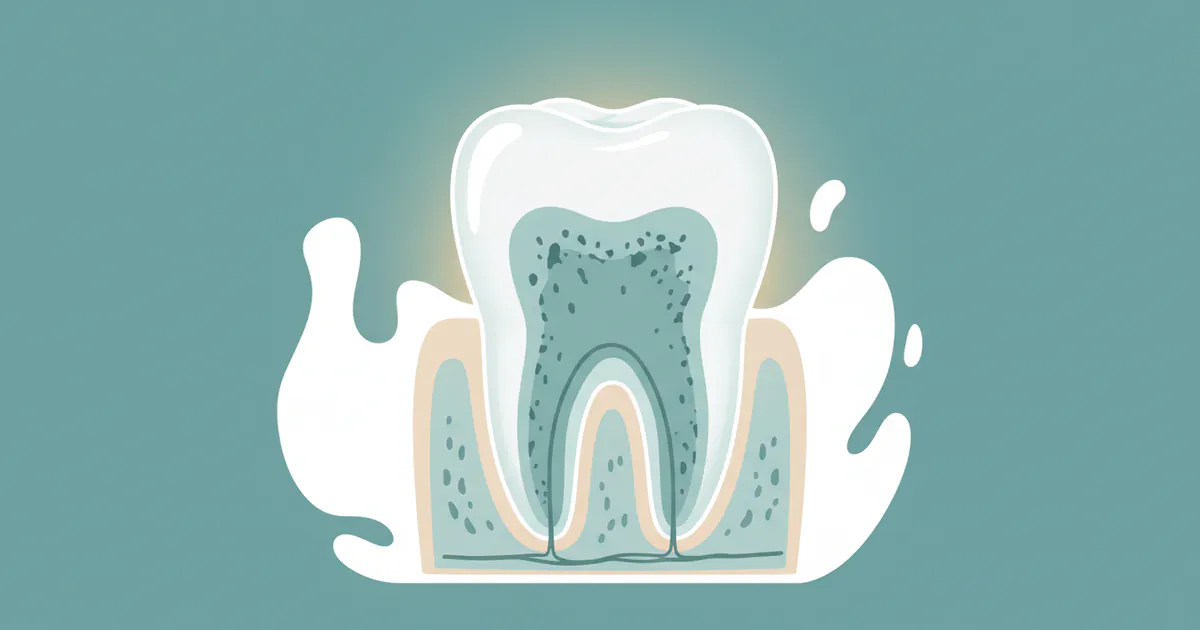
Dentinogenesis imperfecta (DI) is a hereditary developmental disturbance of dentin. To fully grasp its impact, it's essential to understand the basic structure of a tooth. Each tooth is composed of four main tissues: the outer enamel (the hardest substance in the human body), the underlying dentin (a bone-like tissue that makes up the bulk of the tooth), the pulp (the innermost soft tissue containing nerves and blood vessels), and cementum (which covers the tooth root). In healthy teeth, the enamel provides a strong protective layer, and the dentin provides support and resilience.
With dentinogenesis imperfecta, the primary defect lies in the dentin. The collagen fibers and mineral content within the dentin are abnormally formed, making the tissue softer and less stable than it should be. This compromised dentin leads to a cascade of problems. The enamel, which relies on the strong foundation of dentin for support, often fractures or chips away easily, especially under chewing forces. This exposes the underlying, poorly mineralized dentin, which then wears down rapidly. The pulp chambers and root canals, which house the tooth's vital tissues, may also be affected, often becoming progressively smaller or completely obliterated (closed off) over time due to continued dentin formation (termed "obliterated pulp chambers"). This abnormality is evident in both primary (baby) and permanent teeth.
DI is classified as a mesodermal anomaly because dentin is derived from the mesoderm, one of the primary germ layers in embryonic development. It is distinct from other conditions like amelogenesis imperfecta, which affects only the enamel. The genetic nature of DI means it is passed down through families, making early identification and family history crucial for diagnosis and counseling.
Types / Variations
Dentinogenesis imperfecta is broadly categorized into three main types, each with distinct clinical and genetic characteristics. Understanding these variations is crucial for accurate diagnosis and tailored treatment plans.
Dentinogenesis Imperfecta Type I (DI-I)
This type of DI is less common and is almost always associated with osteogenesis imperfecta (OI), often referred to as "brittle bone disease." OI is a systemic connective tissue disorder characterized by fragile bones that fracture easily, blue sclera (the whites of the eyes appear bluish), hearing loss, and joint laxity.
- Genetic Cause: Mutations in the COL1A1 or COL1A2 genes, which are responsible for producing Type I collagen. Type I collagen is abundant in bone, dentin, and other connective tissues, explaining the systemic manifestations.
- Dental Features: Both primary and permanent teeth are affected. Teeth typically appear translucent and opalescent, ranging from blue-gray to amber-brown. The enamel frequently chips away, exposing the soft, abnormally formed dentin, which then wears down quickly. Radiographically (on X-rays), teeth show short roots, bulbous crowns (bell-shaped), constricted cervical necks (the area near the gum line), and partial or total obliteration of the pulp chambers and root canals.
- Systemic Features: Bone fragility (multiple fractures), hearing loss (especially progressive sensorineural hearing loss), blue sclera, hypermobility of joints.
Dentinogenesis Imperfecta Type II (DI-II)
Also known as hereditary opalescent dentin, DI-II is the most common form of dentinogenesis imperfecta and is not associated with any systemic disorder like osteogenesis imperfecta. It primarily affects the dentition.
- Genetic Cause: Mutations in the DSPP gene (Dentin Sialophosphoprotein). This gene provides instructions for making proteins crucial for the mineralization and structure of dentin.
- Dental Features: Clinically and radiographically, the dental features are very similar to those of DI-I. Teeth are opalescent, discolored (blue-gray or amber-brown), translucent, and prone to rapid wear and fracture of enamel. X-rays reveal bulbous crowns, short roots, constricted cervical necks, and early or complete obliteration of pulp chambers and root canals. Both primary and permanent dentitions are affected.
- Systemic Features: None. This type exclusively affects the teeth.
Dentinogenesis Imperfecta Type III (DI-III)
This is the rarest form, often referred to as the "Brandywine type" because it was initially identified in a large, isolated population in Brandywine, Maryland. DI-III is also primarily a dental condition.
- Genetic Cause: Also linked to mutations in the DSPP gene, similar to DI-II, but possibly different mutations or allelic variations lead to a distinct clinical presentation.
- Dental Features: While sharing features with DI-II, DI-III often presents with more severe manifestations. Teeth exhibit the characteristic opalescent appearance and rapid wear. However, a hallmark of DI-III is the presence of "shell teeth" – teeth with extremely large pulp chambers (appearing as hollow shells) and very thin dentin. This differs from the pulp obliteration seen in DI-I and DI-II. Over time, these large pulp chambers can become obliterated. Multiple periapical radiolucencies (areas of bone loss around the root tips indicating infection) and un-erupted teeth are also common.
- Systemic Features: None, exclusively dental.
The distinction between these types is vital for diagnosis, particularly differentiating DI-I from DI-II and DI-III, as the presence of osteogenesis imperfecta has profound implications for a patient's overall health and management.
| Feature | Dentinogenesis Imperfecta Type I | Dentinogenesis Imperfecta Type II | Dentinogenesis Imperfecta Type III |
|---|---|---|---|
| Associated Condition | Osteogenesis Imperfecta (OI) | None | None |
| Genetic Cause | COL1A1 or COL1A2 gene mutations | DSPP gene mutations | DSPP gene mutations (specific variants) |
| Prevalence | Less common (with OI) | Most common form of DI | Rarest form (Brandywine isolate) |
| Clinical Appearance | Opalescent (blue-gray to amber-brown), translucent | Opalescent (blue-gray to amber-brown), translucent | Opalescent, translucent, often severe wear |
| Radiographic Features | Bulbous crowns, constricted cervical necks, short roots, early/complete pulp obliteration | Bulbous crowns, constricted cervical necks, short roots, early/complete pulp obliteration | Bulbous crowns, constricted cervical necks, short roots, "Shell teeth" (very large pulp chambers), multiple periapical radiolucencies |
| Affected Dentition | Primary and Permanent | Primary and Permanent | Primary and Permanent |
Causes / Why It Happens
Dentinogenesis imperfecta is fundamentally a genetic disorder, meaning it results from mutations in specific genes that govern the formation and mineralization of dentin. The inheritance pattern for all types of DI is typically autosomal dominant. This means that only one copy of the mutated gene from one parent is sufficient to cause the condition in a child, regardless of the child's sex. If one parent has DI, there is a 50% chance that each child they have will inherit the condition. While most cases are inherited, new mutations can occasionally occur spontaneously in individuals with no family history, though this is less common.
Genetic Basis
As detailed in the types section, the specific genes involved vary by type:
-
DI Type I: Caused by mutations in the COL1A1 or COL1A2 genes. These genes are crucial for producing alpha-1 and alpha-2 chains of Type I collagen. Type I collagen is the most abundant structural protein in the human body, forming the framework for bones, skin, tendons, ligaments, and importantly, dentin. When these genes are mutated, the collagen produced is either defective or insufficient, leading to fragile bones (osteogenesis imperfecta) and abnormal dentin. The defective collagen impairs the proper mineralization process, resulting in soft, disorganized dentin.
-
DI Type II and Type III: Both are caused by mutations in the DSPP gene (Dentin Sialophosphoprotein). The DSPP gene provides instructions for making a precursor protein that is later processed into two main proteins: dentin sialoprotein (DSP) and dentin phosphoprotein (DPP). These proteins play a critical role in controlling the mineralization process of dentin, promoting the formation of highly organized hydroxyapatite crystals that give dentin its strength and resilience. Mutations in DSPP lead to the production of abnormal or non-functional DSPP proteins. This disrupts the intricate process of dentin formation, resulting in improperly formed and mineralized dentin that is softer, more permeable, and structurally unsound. The specific mutation within the DSPP gene can influence the severity and exact presentation of the condition, explaining the differences between DI-II and DI-III.
In essence, the root cause of dentinogenesis imperfecta is a molecular defect in the building blocks or regulatory proteins of dentin, leading to a tooth structure that cannot withstand normal oral functions.
Signs and Symptoms
Recognizing the signs and symptoms of dentinogenesis imperfecta is critical for early diagnosis and intervention. These manifestations can appear in both primary (baby) teeth and permanent teeth, often becoming noticeable shortly after eruption.
Primary Dental Manifestations:
- Tooth Discoloration: This is often the first and most striking sign. Affected teeth exhibit a characteristic opalescent appearance, often described as a blue-gray or amber-brown hue. This coloration is intrinsic to the dentin and shines through the enamel, making the teeth appear translucent. The color can vary from a uniform tint to a marbled appearance.
- Enamel Fractures and Rapid Wear: The enamel overlying the defective dentin is normal in composition but lacks adequate support. Consequently, the enamel is prone to chipping and fracturing easily, especially on chewing surfaces. Once the enamel is gone, the underlying soft, poorly mineralized dentin wears down at an accelerated rate, leading to severe attrition (wear) of the teeth. This rapid wear can significantly shorten the clinical crown height of teeth.
- Sensitivity: As enamel fractures and dentin wears down, teeth can become highly sensitive to temperature changes (hot/cold), sweet foods, and even air. This is due to the exposure of the dentinal tubules, which transmit stimuli directly to the pulp.
- Short and Bulbous Crowns: The crowns of the teeth often have a distinctive "bulbous" or bell-shaped appearance, with a constriction at the cervical neck (the part of the tooth near the gum line).
- Small or Obliterated Pulp Chambers and Root Canals: On dental X-rays, a characteristic finding is the partial or complete obliteration (closure) of the pulp chambers and root canals. This occurs because the abnormal dentin continues to form throughout the tooth's life, gradually filling these spaces. In DI Type III, "shell teeth" with extremely large pulp chambers are initially seen, which may later become obliterated.
- Shortened or Tapered Roots: The roots of the teeth may appear short, blunt, or slender on radiographs, further reducing their stability in the jawbone. This predisposes teeth to mobility and premature loss.
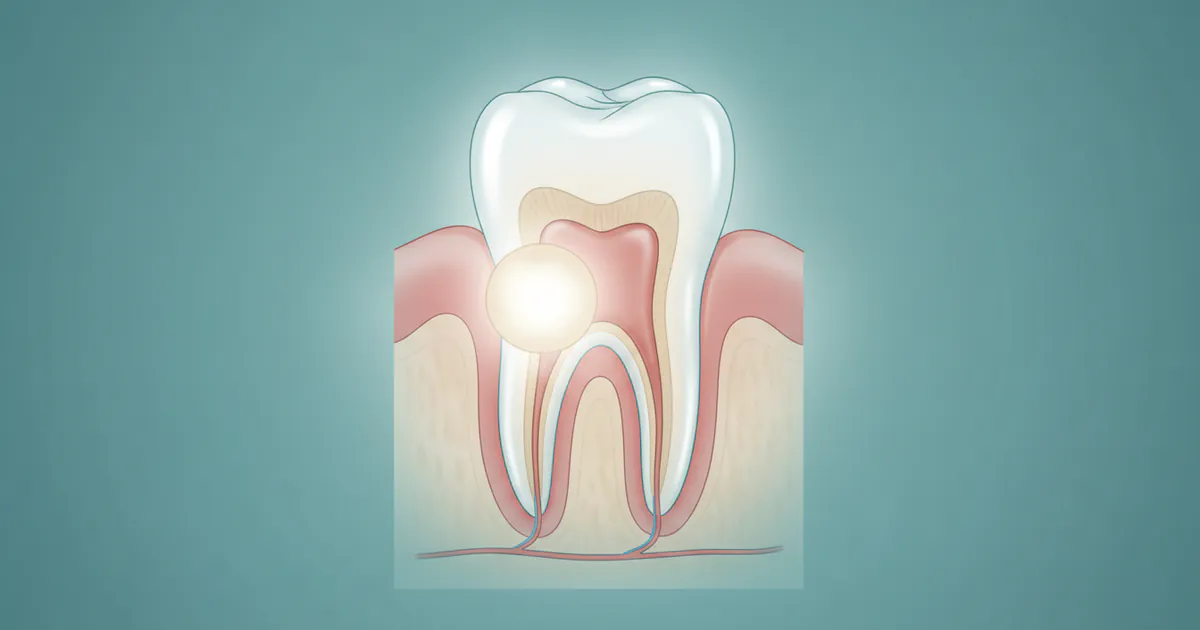
- Increased Susceptibility to Infection: Due to rapid wear and enamel fractures, the underlying dentin and eventually the pulp can become exposed to the oral environment. Even without obvious physical exposure, the compromised dentin structure may allow bacterial penetration. This dramatically increases the risk of pulpitis (inflammation of the pulp) and pulpal necrosis (death of the pulp tissue), which can then lead to periapical infections and the formation of an abscess on gums. An abscess manifests as a painful, swollen bump near the tooth root, often filled with pus, and requires urgent dental attention. Signs of an abscess include severe, throbbing pain, sensitivity to hot/cold, swelling of the gum or face, fever, and a foul taste in the mouth.
- Difficulty with Chewing and Speech: The severe wear and loss of tooth structure can impair the ability to chew food effectively and may also affect speech articulation.
Differentiating from Cavities (How to know if you have a cavity):
While the discoloration and rapid wear in DI can sometimes be mistaken for severe dental decay, there are key differences:
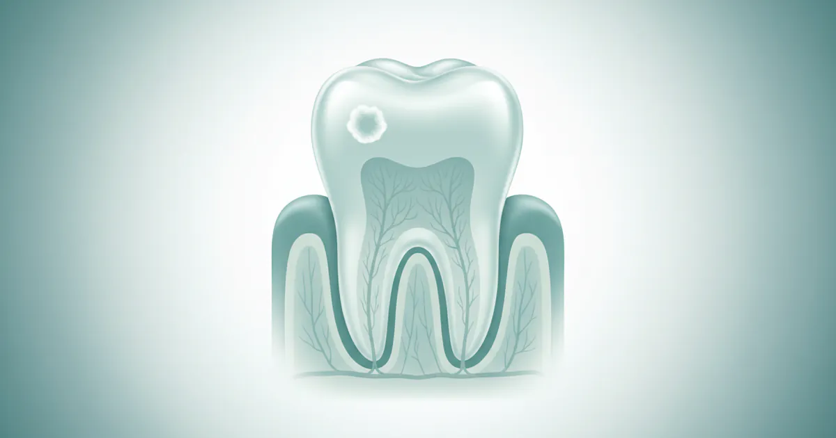
- Cavities (dental caries) are localized areas of damage caused by acid-producing bacteria, typically starting as a small pit or spot that progressively enlarges. They often present as dark spots, holes, or sensitivity in specific areas of a tooth.
- Dentinogenesis imperfecta causes a generalized, intrinsic discoloration and structural weakness affecting most, if not all, teeth. The wear is broad and affects entire chewing surfaces, not just isolated pits. While DI teeth can certainly develop cavities, the primary issue is the inherent defect in the dentin, not bacterial destruction as the initial cause of widespread damage.
- If you suspect you have a cavity due to a dark spot or a hole, it’s crucial to see a dentist. In DI patients, however, the extensive wear might mimic a large cavity, but the underlying issue is systemic. A dentist can differentiate this using clinical examination and X-rays, noting the characteristic radiographic features of DI.
Systemic Manifestations (Specific to DI Type I):
For individuals with DI Type I, the dental symptoms are accompanied by signs of osteogenesis imperfecta (OI):
- Bone Fractures: Frequent bone fractures, often with minimal trauma.
- Blue Sclera: The whites of the eyes appear bluish due to the thinning of the sclera, allowing the underlying choroidal vessels to show through.
- Hearing Loss: Progressive hearing loss, which can be conductive, sensorineural, or mixed.
- Joint Laxity: Hypermobile joints.
Early recognition of any of these signs warrants immediate dental and medical consultation for proper diagnosis and management.
Treatment Options
Managing dentinogenesis imperfecta is a lifelong process focused on protecting the remaining tooth structure, restoring function, improving aesthetics, and preventing complications. Treatment strategies vary depending on the severity of the condition, the patient's age, and the type of DI. The overarching goals are to maintain the integrity of the teeth, prevent further wear, and mitigate pain and infection.
1. Protective Restorations (for preserving tooth structure)
These are typically the first line of defense, especially for young patients, to prevent rapid wear and fracture.

-
Composite Bonding/Resin Restorations:
- Description: Tooth-colored resin material is applied directly to the teeth, sculpted, and hardened with a light. It can be used to cover exposed dentin, repair chips, and build up worn surfaces.
- Pros: Minimally invasive, relatively inexpensive, good aesthetic match, can be done in one appointment.
- Cons: Less durable than crowns, prone to staining, may require frequent repairs or replacement, especially with severe DI.
- Cost: $200 - $600 per tooth (varies by extent).
-
Stainless Steel Crowns (SSCs) for Primary Teeth:
- Description: Pre-formed metal crowns are cemented over primary teeth, providing full coverage and robust protection.
- Pros: Very durable, cost-effective for children, protect teeth from wear and fracture, maintain space for permanent teeth.
- Cons: Aesthetic compromise (silver color), require removal when permanent teeth erupt.
- Cost: $250 - $450 per tooth.
-
Full Coverage Crowns (Permanent Teeth):
- Description: After tooth preparation, a custom-made crown (porcelain-fused-to-metal, all-ceramic, or zirconia) is cemented over the entire tooth.
- Pros: Excellent protection against wear and fracture, durable, highly aesthetic (especially all-ceramic/zirconia).
- Cons: More invasive (requires significant tooth reduction), higher cost, may need replacement over time.
- Cost: $800 - $2,500 per tooth depending on material and location. Zirconia and E-max are often preferred for durability.
-
Veneers:
- Description: Thin, custom-made shells of porcelain or composite material that are bonded to the front surface of teeth.
- Pros: Excellent aesthetic results, can correct discoloration and minor shape irregularities.
- Cons: Primarily aesthetic, may not provide sufficient protection for severely worn or structurally weak teeth, requires some tooth reduction. Not suitable if significant occlusal (biting surface) wear is occurring.
- Cost: $900 - $2,000 per tooth (porcelain).
2. Endodontic Treatment (Pulp Therapy)
Due to pulp obliteration, root canal treatments can be extremely challenging or impossible in DI teeth. However, if an infection arises from pulp exposure, it may be necessary.
- Root Canal Therapy (RCT):
- Description: If the pulp is exposed or infected, and canals are accessible, the infected pulp tissue is removed, and the canal system is cleaned, shaped, and filled.
- Pros: Can save an infected tooth, eliminate pain and infection.
- Cons: Technically challenging in DI teeth due to narrow or obliterated canals, may not be feasible. DI dentin is also more brittle, increasing risk of fracture during treatment.
- Cost: $700 - $1,500 per tooth (varies by tooth type and complexity).
3. Extractions and Prosthetic Replacement (for irreversible cases)
When teeth are too severely damaged, infected beyond repair, or lost due to DI complications.
-
Extractions:
- Description: Surgical removal of the tooth.
- Pros: Eliminates source of pain/infection, necessary if tooth cannot be saved.
- Cons: Loss of natural tooth, requires replacement.
- Cost: $150 - $400 per tooth (simple extraction), $250 - $600+ per tooth (surgical extraction).
-
Dental Implants:
- Description: A titanium screw is surgically placed into the jawbone to replace the root of a missing tooth, over which a crown is placed.
- Pros: Highly durable, mimics natural teeth, preserves bone, excellent aesthetics and function.
- Cons: High cost, surgical procedure, requires sufficient bone density (which might be an issue in DI-I patients due to OI).
- Cost: $3,000 - $6,000 per implant (implant post and abutment, crown extra). Full implant-supported denture could be $20,000 - $50,000.
-
Removable Partial or Complete Dentures:
- Description: Removable appliances that replace multiple missing teeth (partials) or all teeth (complete dentures).
- Pros: Cost-effective solution for multiple missing teeth, restores aesthetics and some function.
- Cons: Less stable and retentive than implants, may affect speech, requires removal for cleaning, can accelerate bone loss over time.
- Cost: $600 - $2,500 for a partial denture, $1,500 - $4,000 for a complete denture (per arch).
-
Fixed Bridges:
- Description: Consists of one or more artificial teeth (pontics) held in place by dental crowns cemented onto adjacent natural teeth (abutments).
- Pros: Fixed solution, good aesthetics, relatively durable.
- Cons: Requires reduction of healthy adjacent teeth, less durable than implants, not suitable if abutment teeth are compromised by DI.
- Cost: $2,000 - $5,000 for a three-unit bridge.
4. Orthodontic Treatment
Orthodontic treatment (braces or aligners) may be considered to correct malocclusion, but it must be approached with caution due to the compromised root structure and bone quality (especially in DI-I). Forces must be light and meticulously monitored to prevent root resorption or tooth loss.
Pro Tip: For children with DI, early intervention with full-coverage stainless steel crowns on primary molars and even anterior teeth can protect them from rapid wear and greatly improve their ability to eat and speak. These provisional crowns can buy time until permanent dentition fully develops and more definitive restorations can be placed.
Comparison of Key Treatment Options for DI
| Treatment Type | Description | Pros | Cons | Average Cost (US) | Longevity |
|---|---|---|---|---|---|
| Composite Bonding | Resin material applied directly to tooth | Minimally invasive, aesthetic, single visit | Less durable, prone to staining, frequent repairs | $200 - $600 per tooth | 2-5 years |
| Stainless Steel Crowns (Primary) | Pre-formed metal cap for baby teeth | Very durable, cost-effective, maintain space | Aesthetic compromise, temporary | $250 - $450 per tooth | 5-10 years (until exfoliation) |
| Full Coverage Crowns (Permanent) | Custom-made cap (porcelain, zirconia) over tooth | Excellent protection, durable, highly aesthetic | Invasive, higher cost, requires significant tooth reduction | $800 - $2,500 per tooth | 10-15+ years |
| Veneers | Thin shell bonded to front of tooth | Excellent aesthetics, corrects discoloration | Primarily aesthetic, less protective for severe wear | $900 - $2,000 per tooth | 7-15 years |
| Dental Implants | Titanium post in jawbone with crown | Highly durable, preserves bone, natural feel | High cost, surgical, requires good bone density | $3,000 - $6,000+ per unit | 20+ years, often lifelong |
| Dentures | Removable appliance for missing teeth | Cost-effective for multiple teeth, restores aesthetics | Less stable, can affect speech, bone loss over time | $600 - $4,000 per arch | 5-10 years (requires relining) |
Step-by-Step Process
Managing dentinogenesis imperfecta often involves a multi-stage, long-term treatment plan. Here’s a generalized step-by-step process you can expect:
Step 1: Initial Diagnosis and Assessment (Early Childhood)
- Clinical Examination: The dentist will observe the characteristic tooth discoloration (opalescent, blue-gray, or amber-brown), evidence of rapid wear, and enamel chipping.
- Radiographic Examination (X-rays): Dental X-rays are crucial. They reveal the typical features of DI: bulbous crowns, constricted cervical necks, short or tapered roots, and the partial or complete obliteration of pulp chambers and root canals. In DI-III, large pulp chambers ("shell teeth") may be evident.
- Family History: A detailed family history helps identify the autosomal dominant inheritance pattern.
- Medical History: Especially important for DI Type I, to assess for signs of osteogenesis imperfecta (history of fractures, blue sclera, hearing issues).
- Genetic Testing: May be recommended to confirm the specific type of DI and identify the causative gene mutation, which can be important for genetic counseling.
- Referral: If DI Type I is suspected, referral to a medical geneticist or an orthopedic specialist is essential.
Step 2: Treatment Planning and Discussion
- Based on the diagnosis, the dentist will develop a comprehensive, individualized treatment plan. This plan will consider the patient's age, severity of DI, presence of primary vs. permanent teeth, and specific needs.
- The dentist will discuss all available options, their pros and cons, costs, and expected longevity.
- Patient and parent education is critical to ensure a clear understanding of the condition and the long-term commitment required for management.
Step 3: Protecting Primary Dentition (Childhood)
- Early Intervention: As soon as primary teeth erupt, protection is paramount.
- Stainless Steel Crowns (SSCs): These are typically placed on posterior (back) primary teeth to prevent wear and fracture, maintaining chewing function and space for permanent teeth.
- Composite Resins: May be used on anterior (front) primary teeth for aesthetic improvement and some protection.
- Monitoring: Regular check-ups (every 3-6 months) are essential to monitor wear, detect new fractures, and address any signs of infection, such as an abscess on gums.
Step 4: Managing Mixed Dentition and Erupting Permanent Teeth
- Space Management: If primary teeth are lost prematurely, space maintainers may be necessary to prevent adjacent teeth from shifting and blocking the eruption of permanent teeth.
- Interceptive Orthodontics: In some cases, light orthodontic intervention may be considered, but with extreme caution due to fragile roots.
- Monitoring Erupting Permanent Teeth: As permanent teeth erupt, they are immediately vulnerable. The dentist will evaluate each tooth for the need for protective restorations.
Step 5: Restoring Permanent Dentition (Adolescence and Adulthood)
- Full Coverage Crowns: This is often the definitive treatment for permanent teeth. Teeth are prepared, and custom-made crowns (porcelain-fused-to-metal, all-ceramic, or zirconia) are fabricated and cemented into place. This process usually takes two appointments:
- Appointment 1: Local anesthesia is administered. The tooth is gently prepared by removing a small amount of enamel and dentin to create space for the crown. Impressions are taken, and a temporary crown is placed.
- Appointment 2: The temporary crown is removed, and the permanent crown is fitted, adjusted for bite and aesthetics, and then permanently cemented.
- Veneers: May be an option for anterior teeth if wear is not severe and the primary concern is aesthetics.
- Composite Restorations: Can be used for smaller repairs or as interim solutions.
- Endodontic Treatment (if necessary): If a pulp infection occurs and canals are accessible, root canal therapy is performed before placing a crown.
- Orthodontics: If needed, usually performed before final restorative work.
Step 6: Addressing Tooth Loss (Adulthood)
- Despite best efforts, some teeth may be lost prematurely.
- Dental Implants: If bone quality is adequate, implants are often the preferred option for replacing missing permanent teeth due to their stability and bone-preserving qualities.
- Fixed Bridges or Removable Dentures: These are alternative options for replacing missing teeth, depending on the number of teeth lost and the patient's budget and preferences.
Step 7: Lifelong Maintenance and Follow-up
- Regular Dental Check-ups: Patients with DI require diligent follow-up care, typically every 3-6 months.
- Oral Hygiene Instruction: Meticulous home care is crucial to prevent secondary decay and gum disease.
- Nightguards/Occlusal Splints: May be recommended to protect restorations and natural teeth from nocturnal grinding (bruxism) or clenching.
- Repair and Replacement: Restorations will need repair or replacement over time due to normal wear and tear or the underlying fragility of the teeth.
Cost and Insurance
The financial burden of managing dentinogenesis imperfecta can be substantial due to the need for extensive and lifelong dental care. Costs vary significantly based on the type of treatment, the number of teeth involved, the materials used, the complexity of the case, and the geographic location within the US.

Average US Price Ranges by Treatment Type:
- Initial Diagnosis & Consultation: $50 - $250
- Dental X-rays (Full Mouth Series): $100 - $250
- Composite Bonding/Fillings: $200 - $600 per tooth
- Stainless Steel Crowns (Primary Teeth): $250 - $450 per tooth
- Porcelain/Ceramic Crowns (Permanent Teeth):
- Anterior (Front): $800 - $1,800 per tooth
- Posterior (Back): $1,000 - $2,500 per tooth (often higher for molars)
- Zirconia Crowns: $1,200 - $2,500 per tooth
- Porcelain Veneers: $900 - $2,000 per tooth
- Root Canal Therapy: $700 - $1,500 per tooth (varies by tooth type)
- Simple Extraction: $150 - $400 per tooth
- Surgical Extraction: $250 - $600+ per tooth
- Dental Implant (Post & Abutment only): $3,000 - $6,000 per implant (crown additional)
- Implant Crown: $1,000 - $2,500
- Partial Denture: $600 - $2,500 per arch
- Complete Denture: $1,500 - $4,000 per arch
- Full Mouth Rehabilitation (extensive crowns/implants): Can range from $20,000 to $60,000 or more, depending on complexity and the number of teeth needing full restoration.
US Price Ranges by Region (General Estimates):
- Low Cost Regions (e.g., Midwest, Southern States): Often on the lower end of the ranges listed above.
- Mid Cost Regions (e.g., Mountain West, some Northeast): Average costs.
- High Cost Regions (e.g., Major Metropolitan areas, California, Northeast coastal cities): Often on the higher end, with some procedures exceeding the ranges.
Insurance Coverage Details:
Navigating insurance for DI treatment can be complex:
-
Dental Insurance:
- Most standard dental insurance plans have annual maximums, typically ranging from $1,000 to $2,500 per year. For extensive conditions like DI, these maximums are quickly met, leaving significant out-of-pocket expenses.
- Coverage for major procedures like crowns, implants, and dentures usually falls into the "major restorative" category, often covered at 50-70% after deductibles are met. Preventive and basic services (exams, X-rays, cleanings, fillings) are usually covered at a higher percentage (80-100%).
- Many plans have waiting periods for major services (e.g., 6-12 months).
- Pre-authorization is almost always required for major procedures, where the dentist submits the treatment plan to the insurance company for approval and an estimate of coverage.
-
Medical Insurance:
- Since DI Type I is a systemic condition associated with osteogenesis imperfecta, some aspects of its management, especially diagnostics (e.g., genetic testing) or consultations with medical specialists, might be covered by medical insurance.
- However, the dental treatments themselves are almost universally classified as dental services and typically fall under dental insurance plans.
- For children, some state-specific Medicaid or CHIP (Children's Health Insurance Program) plans may offer more comprehensive dental benefits for special healthcare needs, but coverage varies widely.
Payment Plans and Financing Options:
Given the high costs, many dental offices offer or partner with financing solutions:
- In-Office Payment Plans: Some practices allow patients to pay in installments over several months, often without interest, especially for extensive treatment plans.
- Third-Party Medical Financing: Companies like CareCredit or LendingClub offer specialized healthcare credit cards with various payment plans, including deferred interest options for a specific period (e.g., 6, 12, 18 months) or fixed interest plans for longer terms.
- Health Savings Accounts (HSAs) and Flexible Spending Accounts (FSAs): These tax-advantaged accounts allow individuals to save and pay for qualified medical and dental expenses with pre-tax dollars, significantly reducing the out-of-pocket burden.
Pro Tip: For children requiring extensive care, inquire about dental schools or university clinics. They often provide high-quality care at a reduced cost, as treatments are performed by supervised students or residents. They may also be more experienced with complex cases like DI.
Recovery and Aftercare
Recovery and aftercare for dentinogenesis imperfecta patients are ongoing processes, rather than a one-time event, given the chronic nature of the condition. The goal is to maximize the longevity of restorations, prevent complications, and maintain overall oral health.
Post-Treatment Care for Restorations:
- Pain Management: Following procedures like crown placement or extractions, mild discomfort, sensitivity, or swelling is common. Over-the-counter pain relievers (e.g., ibuprofen, acetaminophen) are usually sufficient. For more invasive procedures, a dentist may prescribe stronger medication.
- Dietary Modifications:
- Immediately After: Stick to soft foods for a few days, especially after crown cementation or extractions. Avoid very hot or cold foods if sensitivity is present.
- Long-Term: Even with restorations, DI teeth (and the restorations themselves) can be vulnerable. Avoid extremely hard, sticky, or chewy foods that could dislodge crowns or cause fractures. Be mindful of ice chewing or using teeth as tools.
- Oral Hygiene:
- Brushing: Brush thoroughly but gently twice a day with a soft-bristled toothbrush and fluoride toothpaste. Electric toothbrushes can be very effective.
- Flossing: Floss daily to clean between teeth and around restorations. Water flossers can be a helpful adjunct, especially around crowns and bridges.
- Antimicrobial Mouthwash: A non-alcoholic, antimicrobial mouthwash may be recommended by your dentist to reduce bacteria and maintain gum health.
Long-Term Aftercare and Maintenance:
- Regular Dental Check-ups: Due to the inherent fragility of DI teeth and the potential for new complications, frequent dental visits are paramount. Typically, patients require check-ups every 3-6 months to:
- Monitor the integrity of existing restorations.
- Check for new areas of wear, fracture, or sensitivity.
- Screen for signs of gum disease or secondary decay.
- Evaluate for the development of abscess on gums or other infections.
- Ensure proper occlusion (bite) and make adjustments as needed.
- Fluoride Therapy: Professional fluoride applications or prescription-strength fluoride toothpastes/rinses may be recommended to strengthen enamel (where present) and make teeth more resistant to decay.
- Protective Appliances (Nightguards/Occlusal Splints): Many individuals, especially those with significant restorative work, benefit from wearing a custom-fitted nightguard. This appliance protects teeth and restorations from the damaging forces of bruxism (teeth grinding) or clenching, which can accelerate wear and fracture in DI patients.
- Address Sensitivity: If sensitivity persists, your dentist might recommend specific desensitizing toothpastes or in-office treatments.
- Follow-up with Specialists: If DI Type I is present, regular follow-ups with medical specialists (e.g., orthopedic surgeon, audiologist) for osteogenesis imperfecta are essential.
Pro Tip: A consistent, gentle oral hygiene routine is crucial. While DI teeth are structurally compromised, good hygiene can prevent secondary issues like cavities and gum disease, prolonging the life of your natural teeth and restorations. Avoid abrasive toothpastes that could further damage exposed dentin.
Prevention
It's important to clarify that dentinogenesis imperfecta itself cannot be prevented because it is a genetic condition. However, preventing the complications and progression of tooth damage is a cornerstone of managing DI. Early diagnosis and proactive intervention are the most effective "preventative" strategies.
1. Genetic Counseling:
- For families with a history of DI, genetic counseling is highly recommended.
- Counselors can explain the inheritance pattern (autosomal dominant), the risk of passing the condition to future children (50% chance for each child if one parent is affected), and discuss options like prenatal diagnosis or preimplantation genetic diagnosis (PGD) for in-vitro fertilization (IVF).
- Understanding the genetic basis helps families make informed decisions.
2. Early Diagnosis and Intervention:
- This is the most critical aspect of preventing severe complications. Children with DI should see a pediatric dentist as soon as their first teeth erupt, or if any signs of discoloration or wear are noted.
- Preventative Restorations: Placing full-coverage restorations (like stainless steel crowns on primary molars) as soon as possible, often before significant wear occurs, is crucial to protect the fragile dentin and enamel from fracture and attrition. This prevents pulp exposure and subsequent infection, which can lead to an abscess on gums.
- Sealants: While enamel often chips off, if intact enamel is present on permanent molars, dental sealants can provide a protective barrier in pits and fissures against decay, especially as the underlying dentin is weaker.
3. Dietary Guidance:
- A diet low in acidic and sugary foods and drinks helps prevent cavities, which can be particularly devastating for teeth already compromised by DI.
- Avoiding excessively hard or sticky foods can reduce the risk of enamel fractures and damage to restorations.
4. Oral Hygiene Education:
- Thorough and consistent daily oral hygiene (brushing twice a day, flossing daily) is essential to prevent secondary dental caries (cavities) and periodontal (gum) disease. While DI is not a cavity, teeth with weakened enamel and dentin are highly susceptible to decay once bacteria gain access. This helps prevent situations that might cause you to wonder how to know if you have a cavity when the problem might be widespread decay accelerated by DI.
5. Regular Dental Visits:
- Frequent check-ups (every 3-6 months) allow the dentist to monitor the condition of the teeth and restorations, identify problems early, and intervene before they become severe. This includes promptly addressing any signs of early decay or infection to prevent an abscess on gums.

6. Mouthguards:
- For individuals involved in contact sports, a custom-fitted athletic mouthguard is vital to protect teeth from trauma, which can easily fracture DI-affected teeth.
- Nightguards (occlusal splints) are also crucial to prevent damage from nocturnal bruxism (grinding) or clenching.
By proactively managing the condition and adopting these preventative strategies, individuals with dentinogenesis imperfecta can significantly improve their long-term oral health and quality of life.
Risks and Complications
Despite comprehensive treatment, individuals with dentinogenesis imperfecta face several risks and potential complications due to the inherent structural weakness of their teeth.
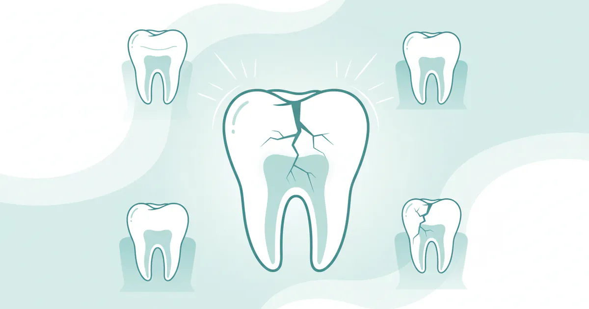
- Rapid Tooth Wear and Attrition: This is a hallmark of DI. Enamel fractures easily, exposing the soft, poorly mineralized dentin, which then wears away rapidly. This leads to severe shortening of the clinical crowns, affecting chewing efficiency and aesthetics.
- Increased Susceptibility to Fractures: Not only is the enamel fragile, but the dentin itself is prone to fracture, particularly the roots. This can lead to tooth loss or make restorative treatment more challenging.
- Pulpal Necrosis and Infection: The rapid wear and enamel loss can lead to premature pulp exposure. Even without gross exposure, the compromised dentin structure may be more permeable to bacteria, leading to inflammation and death of the pulp tissue (pulpal necrosis). This often results in a periapical infection, manifesting as a dental abscess on gums. Signs include persistent pain, swelling, sensitivity to pressure, and sometimes a visible pus-filled bump near the root. Managing these infections can be difficult due to the often-obliterated root canals.
- Early Tooth Loss: Due to severe wear, fractures, root problems, and recurrent infections, premature loss of both primary and permanent teeth is a common complication, even with diligent care.
- Malocclusion and Bite Problems: The loss of tooth structure can disrupt the bite, leading to malocclusion (improper alignment of upper and lower teeth), which can further exacerbate wear and jaw joint issues.
- Periodontal Disease: While not directly caused by DI, the altered tooth structure and complex restorations can sometimes make oral hygiene more challenging, increasing the risk of gum inflammation (gingivitis) and gum disease (periodontitis).
- Aesthetic and Psychological Impact: The discolored, worn, and often fractured appearance of DI teeth can have a significant negative impact on an individual's self-esteem, confidence, and social interactions, especially during childhood and adolescence.
- Challenges with Dental Procedures:
- Root Canal Treatment: The obliterated or severely narrowed pulp chambers and root canals make endodontic therapy extremely challenging, if not impossible, for many DI teeth.
- Restoration Retention: The abnormal dentin and short roots can make it difficult for crowns and other restorations to bond securely or achieve stable retention, potentially leading to debonding or failure.
- Orthodontic Treatment: In DI-I particularly, bone fragility and abnormal tooth roots make orthodontic tooth movement risky, increasing the chances of root resorption or tooth mobility.
- Systemic Complications (DI Type I only): For those with DI Type I, the concurrent osteogenesis imperfecta carries risks of:
- Frequent bone fractures throughout the body.
- Progressive hearing loss.
- Skeletal deformities.
Effective management aims to mitigate these risks, but it's important for patients and their families to be aware of the potential challenges that may arise throughout their lives.
Children / Pediatric Considerations
Dentinogenesis imperfecta often presents in early childhood with the eruption of primary teeth, making pediatric dental care paramount. Early intervention is not just beneficial; it's critical to mitigate the severe consequences of this condition.
Early Diagnosis and Monitoring:
- First Dental Visit: A child with suspected DI should have their first dental visit as soon as the initial primary teeth erupt, or by age one, following ADA recommendations.
- Clinical Signs: Pediatric dentists are trained to recognize the characteristic blue-gray or amber-brown opalescence and rapid wear of the primary teeth.
- Radiographic Evaluation: X-rays are essential to confirm the diagnosis, showing bulbous crowns, constricted cervical necks, short roots, and obliterated pulp chambers, or "shell teeth" in DI-III.
- Parental Education: Parents need comprehensive education about the condition, its hereditary nature, and the lifelong commitment to dental care.
Management of Primary Dentition:
- Prevention of Wear: The primary goal for baby teeth is to protect them from rapid wear and fracture. This is crucial for maintaining chewing function, allowing proper speech development, preserving the vertical dimension of occlusion (the height of the bite), and maintaining space for the permanent teeth.
- Stainless Steel Crowns (SSCs): These are often the treatment of choice for primary molars. They are durable, cost-effective, and provide full coverage, acting as a barrier against wear and exposure of the vulnerable dentin. They also help to maintain proper bite and space.
- Composite Resins: May be used for anterior primary teeth for aesthetic reasons and to provide some protection, though they are less durable than SSCs.
- Space Maintainers: If primary teeth are lost prematurely due to severe wear, fracture, or infection, space maintainers may be necessary to ensure that the permanent teeth can erupt into their correct positions.
Management of Permanent Dentition:
- Proactive Restoration: As permanent teeth erupt, the pediatric dentist will evaluate them for immediate protection. Full-coverage crowns (e.g., all-ceramic or zirconia) are generally recommended for newly erupted permanent molars and premolars to prevent wear and preserve tooth structure.
- Aesthetic Concerns: For anterior permanent teeth, composite bonding or porcelain veneers (when the child is older and growth is complete) can address discoloration and shape.
- Pulp Health: Constant monitoring for pulp exposure and infection is vital. An abscess on gums can develop rapidly in DI teeth due to their compromised structure and susceptibility to bacterial penetration.
- Orthodontic Considerations: Orthodontic treatment requires careful planning and light forces due to the short roots and potential bone fragility (especially in DI Type I). Close collaboration between the orthodontist and the pediatric dentist/general dentist is essential.
Psychological Support:
Children with DI often face aesthetic challenges that can impact their self-esteem and confidence. Support from parents, dentists, and potentially mental health professionals can help them cope with the appearance of their teeth and the extensive dental treatments. Encouraging a positive self-image and focusing on the benefits of treatment are crucial.
Pro Tip: Regular, gentle dental visits from an early age can help normalize the dental experience for children with DI, reducing anxiety and fostering good long-term dental habits.
Cost Breakdown
A detailed breakdown of costs for dentinogenesis imperfecta treatment in the US can help families prepare for the financial implications. These are average ranges, and actual costs can vary significantly.
Average US Costs (Low, Mid, High) for Common Procedures:
| Procedure | Low Range | Mid Range | High Range |
|---|---|---|---|
| Initial Exam & X-rays | $100 | $200 | $350 |
| Composite Restoration (1-surface) | $150 | $250 | $400 |
| Stainless Steel Crown (Primary) | $250 | $350 | $450 |
| Porcelain/Ceramic Crown (Permanent) | $800 | $1,500 | $2,500 |
| Zirconia Crown (Permanent) | $1,200 | $1,800 | $2,500+ |
| Porcelain Veneer | $900 | $1,500 | $2,000 |
| Root Canal (Molar) | $800 | $1,200 | $1,500 |
| Simple Extraction | $150 | $250 | $400 |
| Surgical Extraction | $250 | $400 | $600+ |
| Dental Implant (per unit, incl. crown) | $3,500 | $5,000 | $7,000+ |
| Removable Partial Denture (per arch) | $600 | $1,500 | $2,500 |
| Complete Denture (per arch) | $1,500 | $2,500 | $4,000 |
| Full Mouth Reconstruction | $20,000 | $40,000 | $60,000+ |
With vs. Without Insurance:
- Without Insurance (Cash Pay): You will be responsible for 100% of the listed costs. Dentists may offer a cash discount for upfront payment.
- With Insurance:
- Deductibles: Most dental plans have an annual deductible (e.g., $50-$150) that you must pay out-of-pocket before your insurance begins to cover costs.
- Co-insurance: After meeting your deductible, insurance will cover a percentage of the "allowed amount" for procedures.
- Preventive (Exams, X-rays, Cleanings): Often 80-100% coverage.
- Basic Services (Fillings): Usually 70-80% coverage.
- Major Services (Crowns, Bridges, Implants, Dentures): Typically 50-70% coverage.
- Annual Maximums: The biggest limitation for DI patients is the annual maximum, usually $1,000 - $2,500. This means that once the insurance has paid out this amount within a calendar year, you are responsible for 100% of any further costs for that year. For extensive DI treatment, this maximum is often reached very quickly.
- Lifetime Maximums: Some plans, particularly for orthodontics or certain major services, may have lifetime maximums.
- Pre-authorization: Always get pre-authorization for major procedures to understand your out-of-pocket expenses before treatment begins.
Payment Plans and Financing Options:
- CareCredit/LendingClub: These healthcare credit cards offer promotional financing options, often with 0% APR for a set period (e.g., 6, 12, 18 months) if the balance is paid in full within that time. Longer-term plans with fixed interest rates are also available.
- In-office Payment Plans: Many dental offices are willing to work with patients on extended payment plans, especially for complex treatment. Don't hesitate to discuss this with the office manager.
- Health Savings Accounts (HSAs) & Flexible Spending Accounts (FSAs): If eligible, these accounts allow you to set aside pre-tax money for healthcare expenses, including dental treatment. This can save you a significant amount as the funds are not subject to income tax.
Cost-Saving Tips:
- Early Intervention: Addressing problems early can prevent them from becoming more extensive and costly. Protecting primary teeth with SSCs can prevent premature loss and the need for more complex space management later.
- Max Out Insurance Annually: Plan treatment to utilize your annual maximum each year if multiple procedures are needed, spreading the cost over several years.
- Compare Dentists: Get quotes from a few different dentists or specialists, but prioritize experience with DI over just the lowest price.
- Dental Schools: As mentioned, dental schools or university clinics often provide high-quality care at a reduced cost.
- Negotiate Cash Discounts: If paying out-of-pocket, ask about discounts for upfront payment.
Frequently Asked Questions
What is the life expectancy for someone with dentinogenesis imperfecta?
Dentinogenesis imperfecta does not affect life expectancy. It is a condition specifically impacting dental development, and while it can lead to significant oral health challenges, it does not have systemic health implications that would shorten a lifespan, with the exception of DI Type I, which is associated with osteogenesis imperfecta (brittle bone disease) and can have broader medical impacts.

Is dentinogenesis imperfecta painful?
Yes, DI can be quite painful. The rapid wear and enamel fractures expose the sensitive dentin, leading to increased sensitivity to temperature changes and pressure. Furthermore, the pulp tissue within DI teeth is highly susceptible to infection, which can cause severe pain, swelling, and the formation of a dental abscess on gums. Proactive treatment is essential to manage and prevent pain.
Can dentinogenesis imperfecta be cured?
No, dentinogenesis imperfecta is a genetic condition, and there is no cure. The focus of management is on protecting the existing tooth structure, preventing further damage, restoring function and aesthetics, and managing complications. It requires lifelong dental care and maintenance to preserve the teeth for as long as possible.
What causes an abscess on gums in people with DI?
An abscess on gums in DI patients typically occurs due to pulpal infection. The weak dentin and fragile enamel in DI teeth make them highly prone to rapid wear and fracture, which can expose the pulp chamber to bacteria. Even without direct exposure, the abnormal dentin may be more permeable, allowing bacteria to reach and infect the pulp, leading to its necrosis and subsequent infection of the surrounding bone and gum tissue.
How is dentinogenesis imperfecta diagnosed?
DI is diagnosed through a combination of clinical examination, radiographic (X-ray) analysis, and family history. Clinically, characteristic tooth discoloration (opalescent, blue-gray/amber-brown) and rapid wear are observed. X-rays reveal specific features like bulbous crowns, constricted cervical necks, short roots, and obliterated pulp chambers. Genetic testing can confirm the specific gene mutation, especially useful for genetic counseling.
What is the difference between dentinogenesis imperfecta and amelogenesis imperfecta?
Dentinogenesis imperfecta affects the dentin, the layer beneath the enamel, leading to soft, discolored, and easily worn teeth. Amelogenesis imperfecta, on the other hand, affects only the enamel, causing it to be hypoplastic (thin), hypomineralized (soft), or hypomature (chalky), but the underlying dentin is normal. Both are genetic conditions but affect different tooth tissues.
How long does treatment for DI typically take?
Treatment for DI is a lifelong process. Initial protective restorations in childhood may take a few appointments. As permanent teeth erupt, individual crowns can take 2 appointments per tooth. Full mouth rehabilitation, if necessary, can involve multiple phases over months or even years. Maintenance and replacements will be ongoing throughout life.
Are teeth with DI more prone to cavities?
Yes, while DI itself is not a cavity, teeth affected by DI are highly prone to secondary dental caries (cavities). The compromised dentin structure and fragile enamel make it easier for bacteria to penetrate once the enamel surface is breached, leading to rapid and extensive decay. This is why diligent oral hygiene and early protective restorations are crucial to prevent situations where you might wonder how to know if you have a cavity.
Can I get dental implants if I have DI?
Dental implants can be a viable option for replacing missing teeth in DI patients, especially DI Type II or III. However, careful assessment is needed. Bone quality might be a concern, particularly in DI Type I patients with osteogenesis imperfecta. The success of implants depends on sufficient bone density and stability, which must be evaluated by an oral surgeon or periodontist.
What are "shell teeth" and what type of DI are they associated with?
"Shell teeth" are a characteristic radiographic feature seen in Dentinogenesis Imperfecta Type III (Brandywine type). They appear as teeth with extremely large pulp chambers and very thin layers of dentin. This gives the tooth a hollow, shell-like appearance. Over time, these large pulp chambers may also become obliterated due to continued abnormal dentin formation.
When to See a Dentist
Given the progressive nature of dentinogenesis imperfecta and the potential for severe complications, knowing when to seek professional dental care is crucial.
Immediate Attention (Emergency) – Red Flags:
- Severe, Persistent Toothache: If you or your child experiences throbbing pain that doesn't subside with over-the-counter pain relievers, especially if it worsens when lying down or with hot/cold stimuli. This could indicate a serious pulp infection or a developing abscess on gums.
- Swelling of Gums, Face, or Jaw: Any localized swelling, particularly near a tooth, is a strong indicator of infection. If the swelling is spreading to the face or neck, it is a medical emergency requiring immediate attention from a dentist or even an emergency room. This is a primary sign of an abscess on gums.
- Pus Drainage: A foul taste in the mouth or the visible discharge of pus from the gum near a tooth is a definitive sign of an abscess that needs urgent treatment.
- Sudden Tooth Fracture or Loss: If a significant portion of a tooth breaks off, or a tooth becomes suddenly loose or falls out, it requires prompt dental evaluation to prevent further damage or manage pain.
- Difficulty Swallowing or Breathing (with facial swelling): These are critical signs of a severe and spreading infection that could be life-threatening. Seek emergency medical care immediately.
Scheduled Appointment (Routine Care or New Concerns):
- Characteristic Tooth Discoloration: If you notice blue-gray or amber-brown discoloration in your child's newly erupted teeth or your own, schedule a comprehensive dental examination. This is often the first visible sign of dentinogenesis imperfecta.
- Rapid Tooth Wear or Chipping: If teeth are wearing down quickly, appear shorter, or are frequently chipping and fracturing, this warrants an evaluation.
- Increased Tooth Sensitivity: Persistent or increasing sensitivity to hot, cold, or sweets, beyond minor transient sensitivity, should be checked by a dentist.
- Difficulty Chewing or Speaking: If changes in tooth structure are impacting these functions, a dental assessment is needed.
- Routine Check-ups: For individuals diagnosed with DI, regular dental check-ups every 3-6 months are not just routine but a critical component of ongoing management to monitor the condition, maintain restorations, and prevent complications.
- Suspicion of Cavities: If you're observing dark spots, holes, or localized pain that makes you wonder how to know if you have a cavity, even in DI teeth, a dentist needs to evaluate it promptly as DI teeth are very susceptible to rapid decay.
Early diagnosis and consistent management are the keys to preserving oral health and preventing significant pain and complications for those living with dentinogenesis imperfecta. Do not delay in seeking professional dental care if you notice any concerning signs or symptoms.
Frequently Asked Questions
Medically Reviewed Content
This article was written by our dental health editorial team and reviewed for medical accuracy. Our content follows strict editorial guidelines for reliability and trustworthiness.
Medical Disclaimer
This article is for informational purposes only and does not constitute medical advice. Always consult with a qualified dental professional for diagnosis and treatment. Do not delay seeking professional advice because of something you read on this website.
Related Articles
What Is an Abscess
Imagine a throbbing pain deep within your jaw, a persistent ache that keeps you awake at night and makes even the softest foods unbearable. This isn't just a bad toothache; it could be a sign of something much more serious: a dental abscess. Affecting millions of Americans annually, an abscess is an
February 22, 2026
Cracked Tooth Syndrome: Complete Guide
Cracked Tooth Syndrome: Complete Guide Category: Dental Conditions & Diseases
February 22, 2026
Stage 1 Early Cavity: Complete Guide
Did you know that dental cavities are one of the most common chronic diseases globally, affecting both children and adults? In the United States, over 90% of adults have had a cavity, and a significant portion of these begin as a stage 1 early cavity. Often invisible and painless in its nasc
February 22, 2026
Clove Oil for Toothache: Complete Guide
Few experiences are as acutely distressing as a throbbing toothache, often striking at the most inconvenient times, like the middle of the night. It's a pain that can disrupt sleep, concentration, and overall well-being. While modern dentistry offers definitive solutions, many individuals seek immed
February 22, 2026