Basic Tooth Anatomy: Complete Guide
Key Takeaways
- Understanding the intricate architecture of your teeth is more than just academic curiosity; it's a fundamental step towards lifelong oral health. Did you know that over 90% of American adults have had at least one cavity, a condition directly impacting the core structures of a tooth? To truly p
Understanding the intricate architecture of your teeth is more than just academic curiosity; it's a fundamental step towards lifelong oral health. Did you know that over 90% of American adults have had at least one cavity, a condition directly impacting the core structures of a tooth? To truly protect your smile, prevent common dental issues, and make informed decisions about your oral care, a solid grasp of basic tooth anatomy is essential. This comprehensive guide will take you on a detailed journey through the visible and hidden components of your teeth, explaining their functions, how they develop, and what you can do to maintain their strength and vitality. From the outermost protective layers to the delicate inner nerve network, we'll explore every aspect, empowering you with the knowledge to safeguard your dental well-being.
In this article, we'll delve into the various parts of a tooth, differentiate between primary and permanent dentition, examine the types of teeth and their specific roles, and even touch upon the fascinating timeline of tooth development, including insights into toddler and even dog tooth eruption. We'll also cover common issues related to dental anatomy, how they are treated, and crucial preventative measures.
Key Takeaways:
- Comprehensive Structure: Each tooth comprises three main parts – the crown (visible), the root (anchored in bone), and the neck (gingival margin).
- Protective Layers: Enamel (hardest substance in the body) covers the crown, while cementum protects the root, both overlaying the sensitive dentin.
- Living Core: The pulp, located at the center, contains nerves, blood vessels, and connective tissue vital for tooth health and sensation.
- Periodontal Support: The periodontal ligament and alveolar bone anchor the tooth firmly, enabling it to withstand chewing forces.
- Eruption Timelines: Primary teeth typically erupt between 6-33 months; permanent teeth begin around age 6 and continue into early adulthood.
- Preventative Care: Understanding anatomy highlights the importance of consistent oral hygiene (brushing, flossing) and regular dental check-ups to protect these vital structures.
- Restorative Costs: Addressing issues affecting tooth anatomy can range from $75-$250 for a basic filling to $700-$2,000 for a root canal, emphasizing the value of prevention.
What Is It: A Deep Dive into Basic Tooth Anatomy
At first glance, a tooth might seem like a simple, hard structure, but it is, in fact, a marvel of biological engineering. Each tooth is a complex, living organ designed for chewing, speaking, and maintaining the structure of your jaw and face. Understanding its fundamental components is the cornerstone of appreciating oral health.
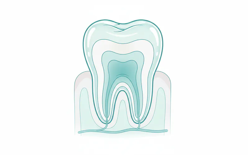
The Major Anatomical Divisions of a Tooth
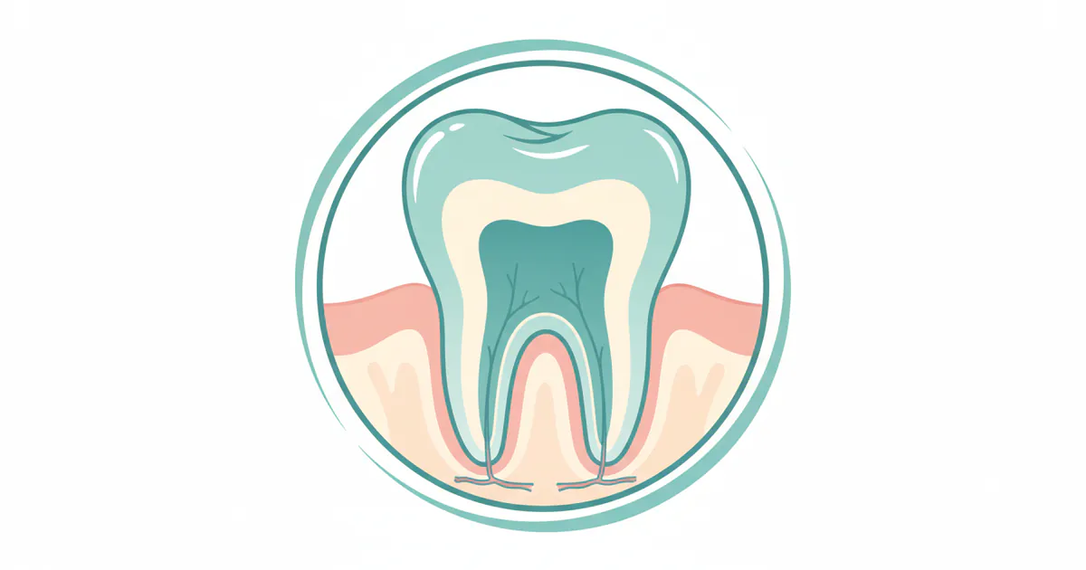
A tooth can be broadly divided into two main parts: the crown and the root(s), with a transitional area known as the neck or cementoenamel junction.
- Crown: This is the visible portion of the tooth, extending above the gum line. Its shape varies depending on the tooth's function (e.g., sharp for incisors, broad for molars). The crown is designed to withstand the immense forces of chewing and biting, making it incredibly durable.
- Root(s): Embedded within the jawbone, the root anchors the tooth firmly in place. While incisors and canines typically have a single root, premolars often have one or two, and molars can have two or three (or rarely, more), providing enhanced stability. These roots are crucial for nutrient supply and sensory feedback.
- Neck (Cervix): This is the narrow junction where the crown meets the root, typically located at the gum line. It's a critical area for oral health, as gum recession can expose the sensitive root surface here.

The Four Main Tissues of a Tooth
Within these divisions lie four specialized tissues, each with unique properties and functions:
- Enamel: The outermost layer of the crown, enamel is the hardest substance in the human body, even tougher than bone. Composed primarily of hydroxyapatite crystals, its incredible strength protects the underlying sensitive tissues from physical and chemical damage during chewing and from acidic foods and drinks. Enamel is translucent and acellular, meaning it cannot regenerate if damaged by cavities or erosion. Its color can range from light yellow to grayish-white, influenced by genetics and thickness.
- Dentin: Located beneath the enamel and cementum, dentin makes up the bulk of the tooth structure. It is a yellowish, bone-like tissue, but softer than enamel. Dentin contains microscopic tubules that run from the pulp to the outer layer, making it sensitive to temperature changes and touch. When enamel is breached, exposed dentin can lead to tooth sensitivity and rapid cavity progression. Dentin is living tissue and can slowly regenerate in response to decay or trauma.
- Pulp: The innermost core of the tooth, the pulp, is a soft, living tissue housed within the pulp chamber (in the crown) and root canals (in the roots). It's a vital part of the tooth, containing nerves, blood vessels, and connective tissue. The nerves provide sensation (pain, pressure, temperature), while the blood vessels supply nutrients and moisture, keeping the dentin vital. When the pulp becomes inflamed or infected due to deep decay or trauma, it can cause severe pain and often necessitates a root canal treatment.
- Cementum: A specialized bone-like tissue that covers the outer surface of the tooth root. Its primary function is to anchor the tooth to the jawbone via the periodontal ligament. Cementum is softer than enamel and dentin and does not contain nerves. It's essential for the stability of the tooth within its socket.
Supporting Structures: The Periodontium
Beyond the tooth itself, several supporting structures collectively known as the periodontium are critical for the tooth's stability and health.
- Periodontal Ligament (PDL): A network of fibrous connective tissues that surrounds the tooth root and connects it to the alveolar bone. The PDL acts as a shock absorber during chewing, distributing forces and preventing damage to the bone. It also contains sensory receptors that provide feedback on biting forces and tooth position.
- Alveolar Bone (Alveolar Process): This is the part of the jawbone that contains the sockets (alveoli) where the teeth are embedded. It provides the primary support for the teeth and is constantly remodeling in response to stresses and forces. Bone loss in this area, often due to periodontal disease, can lead to tooth mobility and eventually tooth loss.
- Gingiva (Gums): The soft tissue that surrounds the teeth and covers the alveolar bone. Healthy gingiva is firm, pink, and fits snugly around the teeth, protecting the underlying bone and root surfaces from bacteria. Inflammation of the gingiva (gingivitis) is the earliest stage of gum disease.
Understanding these foundational components of basic tooth anatomy is the first step in appreciating the complexities of oral health and recognizing the importance of proper dental care.
Types and Variations of Teeth
Humans, like many mammals, have two sets of teeth during their lifetime: primary (deciduous or "baby") teeth and permanent (adult) teeth. Each set comprises different types of teeth, each uniquely shaped and positioned to perform specific functions in the mastication process.
Primary (Deciduous) Teeth
There are typically 20 primary teeth in a child's mouth, 10 in the upper jaw and 10 in the lower. These teeth are smaller, whiter, and have thinner enamel and dentin layers compared to permanent teeth. They play crucial roles in:
- Chewing: Helping children learn to eat solid foods.
- Speech Development: Guiding proper tongue placement for clear speech.
- Space Maintenance: Holding space in the jaw for the developing permanent teeth. Premature loss of primary teeth can lead to crowding of permanent teeth.
The primary dentition includes:
- Incisors (8): Four central and four lateral incisors at the front of the mouth, sharp-edged for biting and cutting food.
- Canines (4): Located next to the lateral incisors, these are pointed teeth for tearing food.
- Molars (8): Two first and two second molars on each side of the jaws, with broad, flat surfaces for grinding food. Primary dentition does not include premolars.
Permanent Teeth
As primary teeth are naturally exfoliated (shed), they are replaced by 32 permanent teeth (including wisdom teeth). These teeth are larger, stronger, and designed to last a lifetime.
The permanent dentition includes:
- Incisors (8): Similar to primary incisors, for biting.
- Canines (4): Pointed teeth for tearing.
- Premolars (8): Also known as bicuspids, these are unique to permanent dentition. They have two cusps (points) and are located between canines and molars, assisting with tearing and grinding.
- Molars (12): Include first, second, and third molars (wisdom teeth). These are the largest teeth, with broad, multi-cusped surfaces for powerful grinding of food. The third molars, or wisdom teeth, are the last to erupt, typically between ages 17 and 25, and may sometimes require extraction due to impaction or lack of space.
Comparison Table: Primary vs. Permanent Teeth
| Feature | Primary (Deciduous) Teeth | Permanent (Adult) Teeth |
|---|---|---|
| Number | 20 | 32 (including wisdom teeth) |
| Color | Whiter, often described as "milk teeth" | More yellowish, darker |
| Size | Smaller | Larger |
| Enamel/Dentin | Thinner layers, less mineralized | Thicker, more mineralized, more durable |
| Pulp Chambers | Relatively larger compared to crown size | Smaller relative to crown size |
| Roots | Shorter, more flared, designed for resorption | Longer, stronger, more divergent |
| Premolars | Absent | Present (8 total) |
| Molars | 8 total (first & second) | 12 total (first, second, third/wisdom) |
| Eruption Age | ~6 months to 3 years | ~6 years to 25 years |
| Purpose | Chewing, speech, space maintenance for permanent teeth | Lifelong mastication, speech, facial structure support |
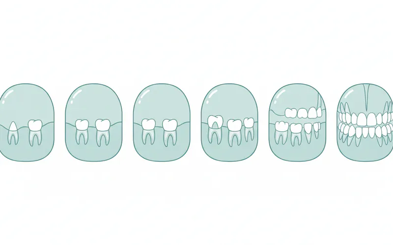
Tooth Development and Eruption
The formation and eruption of teeth (odontogenesis) are complex biological processes that begin even before birth and continue into early adulthood. This developmental timeline is critical for understanding when to expect teeth and when potential issues might arise.
Odontogenesis: The Making of a Tooth
Tooth development starts in the embryonic stage, around the sixth week of gestation. Specialized cells differentiate to form the tooth bud, which then undergoes stages of growth and calcification (hardening) to form the enamel, dentin, and pulp. This process is highly regulated by genetic factors and influenced by maternal health and nutrition.
Tooth Eruption Timelines
Teeth erupt in a generally predictable sequence, though individual variations are common.
Toddler Tooth Eruption Chart (Primary Teeth)
Understanding the toddler tooth eruption chart helps parents anticipate when their child's teeth will emerge and manage associated discomfort.
| Tooth Type | Average Eruption Age (Upper Jaw) | Average Eruption Age (Lower Jaw) |
|---|---|---|
| Central Incisors | 8-12 months | 6-10 months |
| Lateral Incisors | 9-13 months | 10-16 months |
| Canines | 16-22 months | 17-23 months |
| First Molars | 13-19 months | 14-18 months |
| Second Molars | 25-33 months | 23-31 months |
- Pro Tip: While these are averages, a delay of up to 6 months beyond these ranges is often considered normal. Consult your pediatric dentist if you have concerns about your child's tooth development.
Permanent Tooth Eruption Timeline
| Tooth Type | Average Eruption Age (Upper Jaw) | Average Eruption Age (Lower Jaw) |
|---|---|---|
| First Molars | 6-7 years | 6-7 years |
| Central Incisors | 7-8 years | 6-7 years |
| Lateral Incisors | 8-9 years | 7-8 years |
| Canines | 11-12 years | 9-10 years |
| First Premolars | 10-11 years | 10-12 years |
| Second Premolars | 10-12 years | 11-12 years |
| Second Molars | 12-13 years | 11-13 years |
| Third Molars (Wisdom) | 17-21 years (highly variable) | 17-21 years (highly variable) |
Dog Tooth Eruption Chart (Canine Dentition)
While this article focuses on human anatomy, SmilePedia.net recognizes the importance of comprehensive health knowledge. For pet owners, understanding the dog tooth eruption chart can be crucial for monitoring their canine companion's health. Dogs, like humans, have primary (deciduous) and permanent teeth.
Primary (Puppy) Teeth Eruption
Puppies are born without visible teeth.
| Tooth Type | Average Eruption Age (Weeks) | Number of Teeth |
|---|---|---|
| Incisors | 4-6 weeks | 12 |
| Canines | 3-5 weeks | 4 |
| Premolars | 5-6 weeks | 12 |
| Molars | Absent | 0 |
| Total Puppy Teeth | 28 |
Permanent Dog Teeth Eruption
| Tooth Type | Average Eruption Age (Months) | Number of Teeth |
|---|---|---|
| Incisors | 3-5 months | 12 |
| Canines | 4-6 months | 4 |
| Premolars | 4-6 months | 16 |
| Molars | 4-7 months | 10 (upper: 4, lower: 6) |
| Total Permanent Teeth | 42 |
- Note: By 6-7 months, most dogs should have their full set of permanent teeth. Retained primary teeth can be an issue, leading to dental problems and often requiring veterinary extraction.
Maintaining Healthy Tooth Anatomy: Prevention and Care
Understanding the anatomy of your teeth is crucial for preventing common dental problems. Many issues arise when the protective layers or vital internal structures are compromised.
Prevention Strategies
Protecting your basic tooth anatomy involves a combination of consistent home care and professional dental visits.
-
Excellent Oral Hygiene:
- Brushing: Brush at least twice a day for two minutes each time with fluoride toothpaste. Fluoride helps strengthen enamel, making it more resistant to acid attacks and decay, as recommended by the American Dental Association (ADA). Focus on reaching all surfaces of the tooth, including the inner surfaces and chewing surfaces.
- Flossing: Floss daily to remove plaque and food particles from between teeth and under the gum line, areas your toothbrush can't reach. This prevents plaque buildup that can lead to cavities and gum inflammation (gingivitis).
- Mouthwash: Therapeutic mouthwashes can complement brushing and flossing by reducing bacteria and providing extra fluoride, but they are not a substitute for mechanical cleaning.
-
Balanced Diet:
- Limit sugary foods and drinks, which feed oral bacteria that produce acids, leading to enamel erosion and cavities.
- Increase consumption of fruits, vegetables, and dairy products, which provide essential vitamins and minerals for strong teeth and healthy gums. Calcium and Vitamin D are especially important for bone and tooth structure.
- Stay hydrated by drinking plenty of water, especially fluoridated water, which helps rinse away food particles and neutralize acids.
-
Regular Dental Check-ups:
- Visit your dentist at least twice a year for professional cleanings and examinations. These visits allow your dentist to:
- Remove hardened plaque (calculus) that brushing and flossing can't eliminate.
- Detect early signs of cavities, gum disease, or other issues before they become severe, potentially saving you from more extensive and costly treatments.
- Provide fluoride treatments or sealants as needed to protect enamel.
- Assess the health of your supporting structures, like your gums and alveolar bone.
- Visit your dentist at least twice a year for professional cleanings and examinations. These visits allow your dentist to:
-
Protective Measures:
- Mouthguards: If you play contact sports, wear a custom-fitted mouthguard to protect your teeth from trauma that could chip, fracture, or even dislodge them.
- Nightguards: If you grind or clench your teeth (bruxism), a nightguard can protect your enamel from excessive wear and prevent jaw pain.
- Avoid Harmful Habits: Refrain from using your teeth as tools to open packages, bite nails, or chew on ice, as these habits can cause cracks or chips in the enamel.
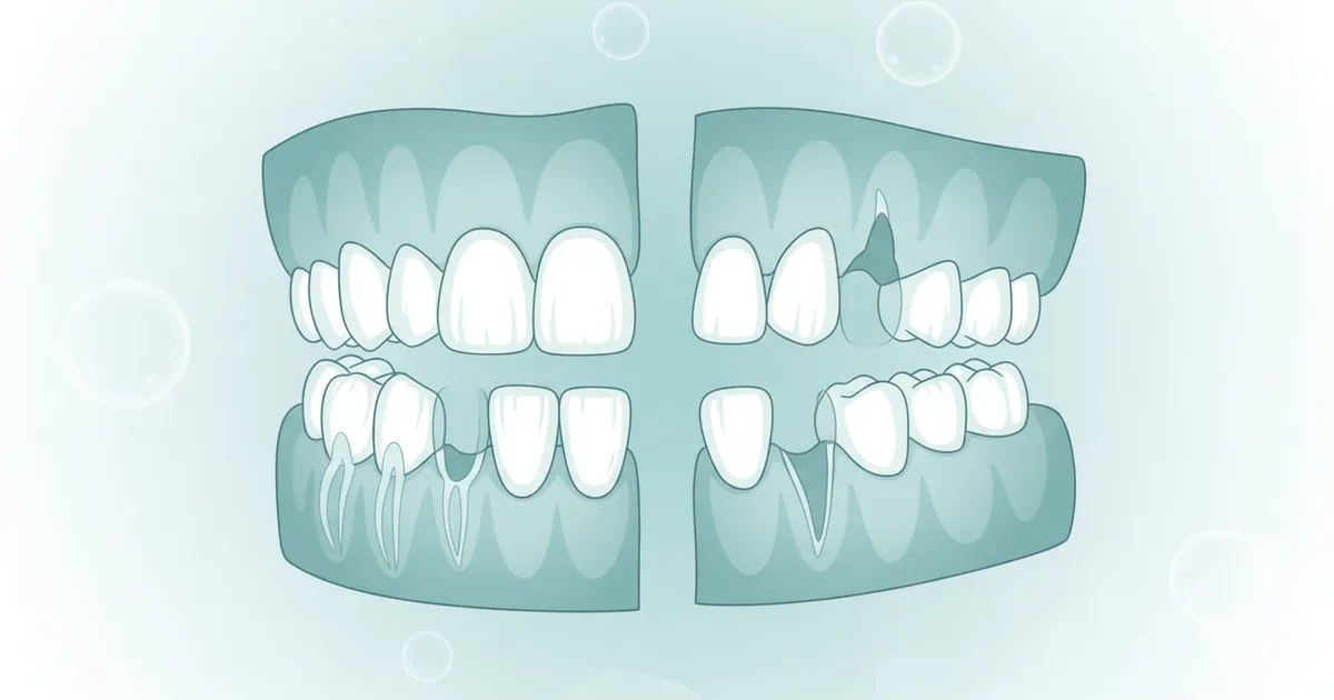
Signs and Symptoms of Compromised Anatomy
While healthy tooth anatomy presents no "symptoms" beyond its normal function, understanding when something is wrong with these structures is key to timely intervention.
- Tooth Sensitivity: A common sign when enamel is worn down or dentin is exposed. This can manifest as a sharp, sudden pain when consuming hot, cold, sweet, or acidic foods and drinks. It can also indicate cavities or gum recession.
- Pain:
- Dull ache: Often associated with early decay reaching dentin or gum inflammation.
- Sharp, throbbing pain: Suggests involvement of the pulp (pulpitis) or an infection within the tooth or surrounding bone.
- Pain on biting/chewing: Can indicate a cracked tooth, a deep cavity, or issues with the periodontal ligament or jawbone.
- Visible Cavities/Stains: Dark spots or holes on the enamel surface are clear signs of decay. Brown or black stains can also indicate enamel demineralization.
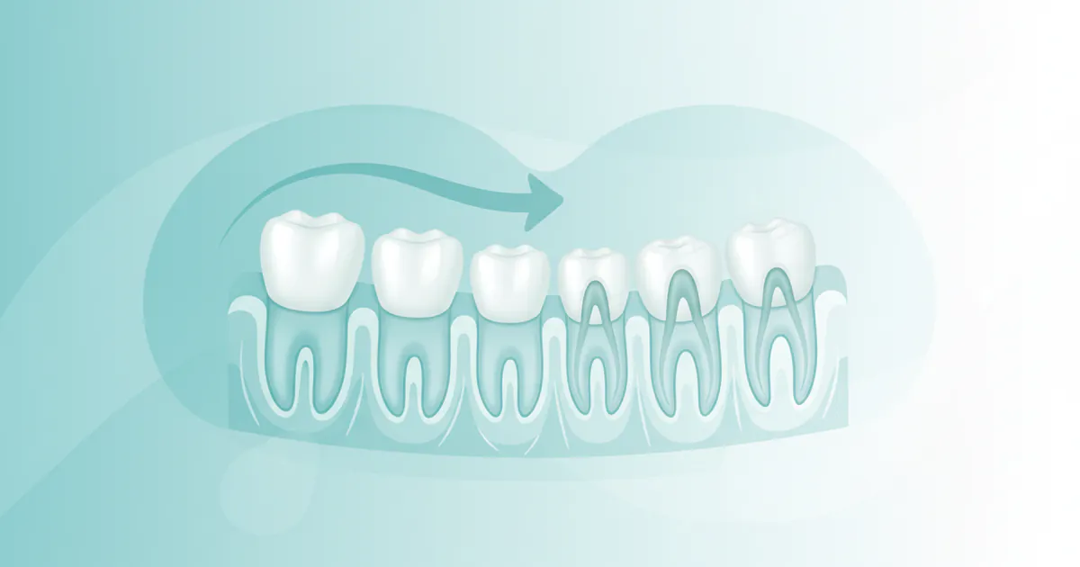
- Swelling/Redness of Gums: Inflammation (gingivitis) or infection (periodontitis) of the gingiva, indicating issues with the supporting structures of the tooth.
- Loose Tooth: Sign of severe gum disease or trauma affecting the periodontal ligament and alveolar bone.
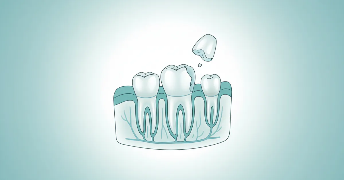
- Chipped/Fractured Tooth: Physical damage to the enamel or dentin, exposing inner layers.
- Bad Breath (Halitosis): Can be a symptom of bacterial buildup, gum disease, or dental decay.
Treatment Options for Anatomical Issues
When basic tooth anatomy is compromised by decay, trauma, or disease, various treatments are available to restore function, prevent further damage, and alleviate pain. The specific treatment depends on which part of the tooth is affected and the severity of the issue.
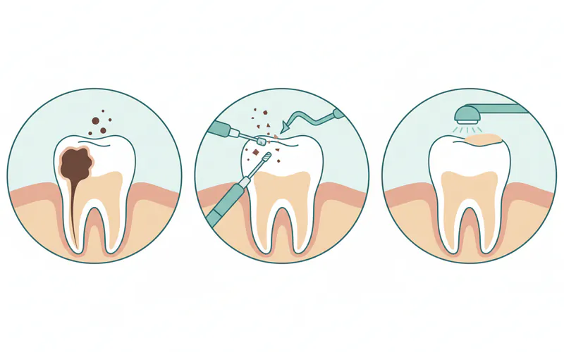
1. Enamel and Dentin Issues (Cavities, Erosion, Chips)
- Dental Fillings: For cavities that have breached the enamel and penetrated the dentin. The decayed portion is removed, and the tooth is filled with materials like composite resin (tooth-colored), amalgam (silver), glass ionomer, or porcelain.
- Pros: Restores tooth structure, prevents further decay, relatively quick procedure.
- Cons: Fillings have a lifespan, amalgam is visible, composite may stain over time.
- Step-by-Step:
- Numbing: Local anesthetic applied to numb the area.
- Decay Removal: Dentist uses a drill to remove decayed tooth structure.
- Cleaning: The cavity is cleaned and disinfected.
- Filling: Restorative material is applied in layers (for composite) and cured with a special light, then shaped and polished.
- Dental Bonding: For minor chips, cracks, or gaps, a tooth-colored resin is applied and sculpted to restore the tooth's appearance.
- Dental Crowns: If a cavity is too large for a filling, or if a tooth is severely cracked or weakened, a crown (a cap covering the entire visible portion of the tooth) may be needed. Crowns are typically made of porcelain, ceramic, metal, or a combination.
2. Pulp Issues (Infection, Inflammation)
- Root Canal Treatment (Endodontics): When the pulp becomes irreversibly inflamed or infected, a root canal is performed to save the tooth.
- Pros: Saves the natural tooth, prevents extraction, resolves pain.
- Cons: Multi-appointment procedure, tooth may become brittle and require a crown.
- Step-by-Step:
- Numbing: Local anesthetic administered.
- Access: A small opening is made in the crown to access the pulp chamber.
- Cleaning: Infected pulp tissue, nerves, and blood vessels are removed from the pulp chamber and root canals.
- Shaping: The canals are cleaned, shaped, and disinfected.
- Filling: The cleaned canals are filled with a biocompatible material (gutta-percha).
- Sealing: The access opening is sealed, and usually a crown is placed for protection.
3. Periodontal Issues (Gums, Periodontal Ligament, Alveolar Bone)
- Scaling and Root Planing: A deep cleaning procedure to remove plaque and calculus from above and below the gum line, smoothing root surfaces to prevent bacterial reattachment. This is often the first line of treatment for gingivitis and early periodontitis.
- Gum Grafts: For gum recession where the root surface is exposed, soft tissue from another part of the mouth (or a donor source) is grafted to cover the exposed root.
- Bone Grafts: In cases of severe bone loss due to periodontal disease or trauma, bone grafting can help regenerate lost alveolar bone, stabilizing the tooth or preparing the site for dental implants.
- Periodontal Surgery: Various surgical procedures may be performed to reduce pocket depths, regenerate lost tissue, or reshape gum tissue.
4. Tooth Loss (Missing Anatomy)
- Dental Implants: A titanium post is surgically placed into the jawbone to act as an artificial tooth root. After healing, a crown, bridge, or denture is attached.
- Pros: Mimics natural tooth, preserves bone, highly durable, long-lasting.
- Cons: Surgical procedure, higher cost, longer treatment time.
- Dental Bridges: Used to replace one or more missing teeth by anchoring artificial teeth to adjacent natural teeth (which are often crowned).
- Dentures: Removable appliances (complete or partial) to replace multiple missing teeth and surrounding tissues.
Cost and Insurance Considerations (US Market)
The cost of dental treatments in the US can vary significantly based on the procedure's complexity, the materials used, the dentist's fees, geographic location, and whether you have dental insurance. It is important to remember that these are general ranges, and obtaining a personalized estimate from your dental provider is always recommended.
Average US Costs by Procedure
| Procedure | Low Range (No Insurance) | Mid Range (No Insurance) | High Range (No Insurance) |
|---|---|---|---|
| Basic Filling (Composite) | $75 | $150 | $250 |
| Amalgam Filling | $50 | $120 | $200 |
| Dental Crown (Porcelain/Ceramic) | $800 | $1,200 | $2,000 |
| Root Canal (Front Tooth) | $700 | $900 | $1,300 |
| Root Canal (Molar) | $1,000 | $1,300 | $2,000 |
| Simple Tooth Extraction | $75 | $200 | $400 |
| Surgical Extraction (e.g., Wisdom) | $200 | $450 | $800 |
| Scaling & Root Planing (per quadrant) | $150 | $300 | $450 |
| Dental Implant (per tooth, implant only) | $1,500 | $2,500 | $3,500 |
| Dental Implant (incl. abutment & crown) | $3,000 | $4,500 | $6,000+ |
- Pro Tip: Costs for procedures like crowns and implants can sometimes be higher in major metropolitan areas or with specialist dentists. Always inquire about all-inclusive pricing for complex treatments.
Insurance Coverage Details
Dental insurance typically categorizes procedures into preventative, basic, and major services, with varying levels of coverage.
- Preventative Care (100% Coverage): Most plans cover 100% of preventative services like routine check-ups, cleanings, and X-rays, recognizing their importance in maintaining tooth anatomy and preventing costly issues.
- Basic Services (70-80% Coverage): Procedures like fillings, simple extractions, and basic periodontal care (e.g., scaling and root planing) are usually covered at 70-80% after you meet your deductible.
- Major Services (50% Coverage): More extensive procedures such as crowns, bridges, dentures, root canals, and oral surgery are typically covered at 50%, often with a deductible and an annual maximum limit (e.g., $1,000-$2,000 per year).
- Orthodontics & Implants: Coverage for these can vary significantly. Some plans offer limited orthodontic benefits for children, while implant coverage is often excluded or very limited.
Payment Plans and Financing Options
For those without adequate insurance or facing high out-of-pocket costs, several financing options exist:
- Payment Plans: Many dental offices offer in-house payment plans, allowing you to pay for treatment in installments.
- Dental Discount Plans: These are not insurance but offer reduced fees for a network of dentists in exchange for an annual membership fee.
- Third-Party Financing (e.g., CareCredit): Medical credit cards that offer promotional interest-free periods if the balance is paid within a specified timeframe.
- Health Savings Accounts (HSAs) & Flexible Spending Accounts (FSAs): If you have these employer-sponsored accounts, you can use pre-tax dollars to pay for dental expenses.
Recovery and Aftercare
Proper aftercare is crucial for the success and longevity of any dental procedure that modifies or restores tooth anatomy. General guidelines include:
- Pain Management: Over-the-counter pain relievers (ibuprofen, acetaminophen) usually suffice. Your dentist may prescribe stronger medication for more invasive procedures.
- Swelling Reduction: Apply ice packs intermittently to the outside of your cheek for the first 24-48 hours.
- Dietary Adjustments: Stick to soft foods for a few days following procedures like fillings, root canals, or extractions. Avoid chewing on the treated side until discomfort subsides.
- Oral Hygiene: Continue gentle brushing and flossing, avoiding the immediate surgical site, if applicable. Your dentist may recommend a special antimicrobial rinse.
- Follow-up Appointments: Attend all scheduled follow-up visits to ensure proper healing and address any concerns.
- Crown Care: After a crown, avoid sticky or hard foods that could dislodge it, and practice good oral hygiene to prevent decay around the crown margin.
- Root Canal Aftercare: It's common for a root canal-treated tooth to be sensitive for a few days. The tooth typically requires a permanent filling and a crown soon after to protect it from fracture.
Risks and Complications
While dental procedures are generally safe, understanding potential risks and complications related to tooth anatomy can help you make informed decisions.
- Fillings: Sensitivity to hot/cold, allergic reaction to material, filling fracture/loss, new decay around the filling.
- Root Canals: Persistent pain, incomplete infection removal (requiring retreatment or extraction), tooth fracture (especially if not crowned), nerve damage (rare).
- Extractions: Dry socket (painful condition after clot dislodges), infection, nerve damage, damage to adjacent teeth, prolonged bleeding.
- Crowns: Sensitivity, nerve damage, allergic reaction, crown loosening or fracture, decay underneath the crown.
- Periodontal Treatment: Sensitivity, gum recession, infection, discomfort.
- Dental Implants: Failure of the implant to integrate with bone, infection, nerve damage, loosening of the crown/abutment.
Adhering to post-treatment instructions and maintaining excellent oral hygiene significantly reduces the risk of complications.
Children / Pediatric Considerations
Children's dental anatomy, though temporary, is fundamental to their oral health development. Pediatric dentistry focuses on nurturing this delicate stage.
The Importance of Primary Teeth
Despite being temporary, primary teeth are crucial. They serve as guides for permanent teeth, ensuring proper alignment and spacing. Early loss of a primary tooth due to severe decay or trauma can lead to shifting of adjacent teeth, causing crowding or misalignment of the permanent teeth erupting later. This underscores why pediatric dentists emphasize treating cavities in baby teeth rather than simply waiting for them to fall out.
Common Pediatric Anatomical Concerns
- Early Childhood Caries (ECC): Often called "baby bottle decay," this severe form of tooth decay can affect infants and toddlers. It primarily impacts the upper front teeth and occurs due to prolonged exposure to sugary liquids. Understanding enamel's vulnerability in primary teeth highlights the need for early intervention.
- Thumb-Sucking/Pacifier Use: Prolonged habits beyond age 2-4 can affect the alignment of erupting teeth and jaw development, impacting occlusion (bite).
- Fluoride Varnish/Sealants: Pediatric dentists often apply fluoride varnish to strengthen enamel and recommend dental sealants for the chewing surfaces of molars, acting as a protective barrier against decay in the pits and grooves that are prone to trapping food and bacteria. These preventative measures leverage understanding of the tooth's surface anatomy.
- Teething Discomfort: During the toddler tooth eruption phase, children may experience discomfort. Parents can offer relief through chilled teething rings, gentle gum massage, or approved pain relievers.
- Dental Trauma: Children are prone to falls and accidents, which can result in chipped, fractured, or dislodged primary or permanent teeth. Prompt dental attention is vital to assess the damage to the underlying tooth anatomy and surrounding structures.
Parents should begin routine dental visits for their child by their first birthday or when their first tooth erupts, as recommended by the ADA and American Academy of Pediatric Dentistry (AAPD). These early visits establish good oral hygiene habits and allow the dentist to monitor the development of the child's teeth and jaws.
Frequently Asked Questions
H3: What is the hardest part of the human tooth?
The hardest part of the human tooth is the enamel, which covers the crown. It is the most mineralized substance in the entire human body, even harder than bone, providing essential protection against chewing forces and acidic attacks.
H3: Can a tooth heal itself if it has a cavity?
No, once a cavity has formed and penetrated the enamel, a tooth cannot heal itself. Enamel is acellular and cannot regenerate. While dentin can partially repair itself, significant decay requires professional dental intervention, such as a filling, to prevent further damage and infection.
H3: How long does a filling last?
The lifespan of a dental filling varies depending on the material, location, and your oral hygiene. Amalgam fillings can last 10-15 years or more, while composite (tooth-colored) fillings typically last 5-10 years. Regular check-ups help your dentist monitor the condition of your fillings.
H3: Is a root canal painful?
Modern root canal treatments are typically performed under local anesthesia, making the procedure itself largely painless, similar to getting a routine filling. Post-procedure, some soreness or mild discomfort is common for a few days, but this is manageable with over-the-counter pain medication. The treatment is designed to relieve the pain caused by the infected pulp.
H3: What is the difference between gums and alveolar bone?
The gums (gingiva) are the soft, pink tissue that surrounds the teeth and covers the alveolar bone. The alveolar bone is the part of the jawbone that holds the tooth sockets, providing structural support to the teeth. Healthy gums protect the bone from bacteria and trauma.
H3: Why do some people need their wisdom teeth removed?
Wisdom teeth (third molars) often need removal because there isn't enough space in the jaw for them to erupt properly. They can become impacted (stuck under the gum or bone), partially erupt, or come in at an angle, leading to pain, infection, damage to adjacent teeth, or cysts.
H3: How much does a dental implant cost compared to a bridge?
A single dental implant with its crown typically costs between $3,000 and $6,000, while a traditional three-unit dental bridge replacing one tooth generally ranges from $2,000 to $5,000. Implants preserve bone and don't affect adjacent teeth, but have a higher upfront cost and longer treatment time.
H3: What causes tooth sensitivity?
Tooth sensitivity typically occurs when the enamel is worn down, exposing the underlying dentin. This can be caused by cavities, gum recession, aggressive brushing, tooth grinding, enamel erosion from acidic foods, or cracked teeth. The exposed dentin tubules allow external stimuli to reach the nerves in the pulp.
H3: Can flossing damage my gums?
No, proper flossing will not damage your gums. In fact, it's essential for gum health. Aggressive or incorrect flossing techniques, however, can irritate or cut the gum tissue. Always use a gentle, C-shaped motion, curving the floss around each tooth and sliding it gently below the gum line.
H3: What role does the periodontal ligament play?
The periodontal ligament (PDL) is a vital fibrous tissue that connects the tooth's root to the alveolar bone. It acts as a shock absorber during chewing, distributes forces, provides sensory feedback for bite pressure, and supplies nutrients to the cementum and bone. It's crucial for tooth stability and function.
When to See a Dentist
Regular dental check-ups are key to maintaining healthy tooth anatomy, but certain signs and symptoms warrant immediate attention. Recognizing these "red flags" can prevent minor issues from becoming major, painful, and costly problems.
Routine Care vs. Red Flags
- Routine Care: Schedule regular check-ups and cleanings every six months (or as recommended by your dentist based on your oral health) even if you feel no pain. This allows for preventative measures and early detection of issues that haven't yet caused symptoms.
- Red Flags (Schedule an appointment promptly):
- Persistent Toothache: Any tooth pain that lasts more than a day or two, especially if it's throbbing, sharp, or awakens you at night. This could indicate a deep cavity, infection, or nerve involvement.
- Swollen or Bleeding Gums: Gums that are consistently red, swollen, tender, or bleed easily during brushing or flossing are signs of gingivitis or periodontitis.
- Extreme Sensitivity: New or worsening sensitivity to hot, cold, sweet, or pressure that impacts your daily life could mean exposed dentin, a new cavity, or a cracked tooth.
- Visible Cavities/Pits: Any dark spots, holes, or pitting on your tooth surfaces.
- Bad Breath (Chronic): Persistent bad breath that doesn't resolve with brushing and flossing can be a sign of gum disease or hidden decay.
- Loose Tooth: Any feeling of a tooth being loose, which could indicate advanced gum disease or trauma.
- Clicking or Painful Jaw: Issues with the temporomandibular joint (TMJ) can sometimes manifest as dental pain.
Emergency vs. Scheduled Appointments
- Emergency (Seek immediate dental care):
- Severe, Uncontrolled Pain: Excruciating toothache that isn't relieved by over-the-counter medication.
- Knocked-Out Tooth (Avulsion): If a permanent tooth is knocked out, attempt to place it back in the socket or keep it in milk/saliva and see a dentist within 30-60 minutes for the best chance of saving it.
- Broken or Chipped Tooth with Severe Pain/Bleeding: Significant trauma to a tooth that causes severe pain, sensitivity, or bleeding.
- Abscess (Facial Swelling): A painful, pus-filled swelling around a tooth or on the gums, especially if accompanied by fever, which indicates a serious infection that can spread.
- Foreign Object Lodged Between Teeth: If you cannot remove it with flossing and it's causing pain or discomfort.
Understanding your basic tooth anatomy empowers you to be an active participant in your oral health journey. By recognizing the intricate structures and their importance, you can better appreciate the need for preventative care and make informed decisions when issues arise, ensuring a healthy, confident smile for years to come.
Frequently Asked Questions
Medically Reviewed Content
This article was written by our dental health editorial team and reviewed for medical accuracy. Our content follows strict editorial guidelines for reliability and trustworthiness.
Medical Disclaimer
This article is for informational purposes only and does not constitute medical advice. Always consult with a qualified dental professional for diagnosis and treatment. Do not delay seeking professional advice because of something you read on this website.
Related Articles
Tooth Eruption Order: Complete Guide
The journey of human teeth, from their initial emergence in infancy to the arrival of wisdom teeth in young adulthood, is a fascinating and crucial aspect of our overall health. Understanding the tooth eruption order is not just a matter of curiosity; it's a vital piece of knowledge that empower
February 23, 2026
Primary Tooth Eruption Sequence: Complete Guide
Did you know that by their third birthday, most children will have a full set of 20 primary teeth? The journey of these first teeth, from their initial appearance to their eventual replacement by permanent teeth, is a remarkable and often challenging milestone for both infants and parents. Understan
February 23, 2026
Canine Tooth Eruption: Complete Guide
Have you ever wondered about the complex process that brings your teeth into alignment, especially the prominent, pointed teeth known as canines? While often overshadowed by discussions of wisdom teeth or incisors, the canine tooth eruption process is a critical stage in your oral development, d
February 23, 2026

Permanent Tooth Eruption Sequence: Complete Guide
Welcome to SmilePedia.net, your trusted resource for comprehensive dental health information. The journey of your permanent teeth emerging into your mouth, known as the permanent tooth eruption sequence, is a fundamental aspect of oral development. Did you know that while primary (baby) teeth ty
February 23, 2026