Early Cavity: Complete Guide
Key Takeaways
- A staggering 91% of American adults over the age of 20 have had a cavity, and 27% currently have untreated tooth decay. These statistics from the CDC highlight a widespread dental health challenge. But what if you could catch a cavity before it becomes a major problem? This is where understandin
A staggering 91% of American adults over the age of 20 have had a cavity, and 27% currently have untreated tooth decay. These statistics from the CDC highlight a widespread dental health challenge. But what if you could catch a cavity before it becomes a major problem? This is where understanding an early cavity becomes crucial. Often unnoticed in its initial stages, an early cavity, also known as incipient caries or demineralization, is the earliest sign of tooth decay. Identifying and treating it promptly can save you from pain, costly procedures, and the potential progression to more severe health issues like a tooth infection. This comprehensive guide from SmilePedia.net will demystify early cavities, exploring their causes, symptoms, diagnosis, a full spectrum of treatment options, costs, prevention strategies, and what to do if you suspect you have one. By empowering you with this knowledge, we aim to help you maintain optimal oral health and preserve your natural smile.
Key Takeaways:
- An early cavity (incipient caries) is the initial stage of tooth decay, characterized by demineralization of enamel, often appearing as a white or light brown spot.
- It is often reversible through remineralization therapies like fluoride varnish or calcium phosphate products if caught before cavitation (a hole) forms.
- Typical treatments for small, cavitated early cavities include dental fillings, which can range from $75 to $400 per tooth depending on material and location.
- Untreated early cavities can progress to severe pain, tooth infection (abscess), and in rare but serious cases, lead to life-threatening systemic infections, making timely treatment essential.
- Prevention is key: diligent oral hygiene, a balanced diet, regular dental check-ups (every 6 months), and professional fluoride treatments are highly effective.
- Costs for preventive treatments like fluoride varnish are typically $25-$75, and sealants $30-$60 per tooth, often covered by insurance for children.
- For suspected early cavities, see a dentist promptly; don't wait for pain to develop, as early intervention dramatically improves outcomes and reduces cost.
``
What It Is / Overview: The Silent Threat of Demineralization
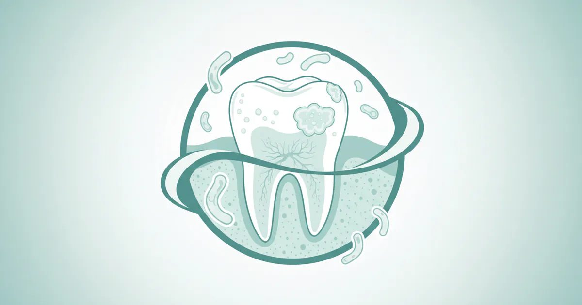
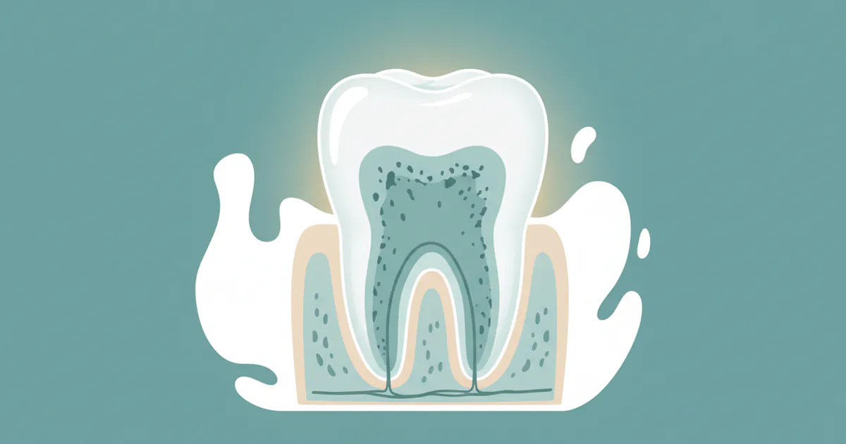
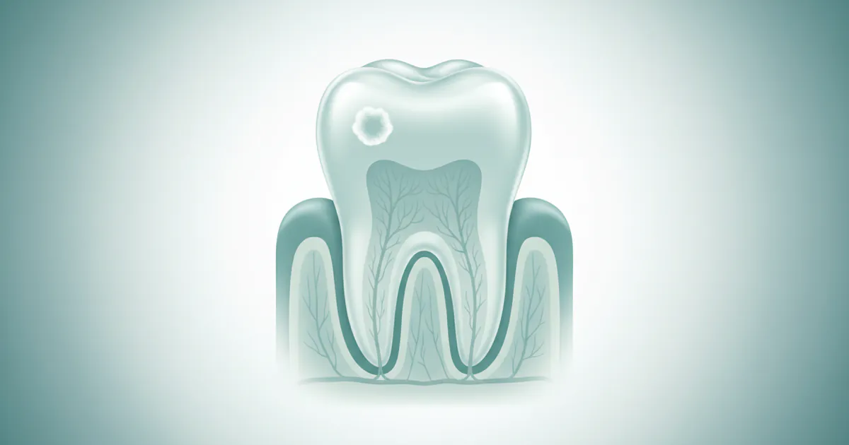
An early cavity, medically termed incipient caries or enamel demineralization, represents the very first stage of tooth decay. It's a localized breakdown of the tooth's outermost layer, the enamel, caused by acids produced by bacteria in your mouth. Unlike a full-blown cavity, which is a visible hole or cavitation in the tooth, an early cavity typically appears as a chalky white spot or a faint discoloration (yellowish or light brown) on the tooth surface. At this stage, the enamel is weakened but often still intact.

Think of your tooth enamel like a hard, protective shell. Throughout the day, a constant tug-of-war happens on its surface:
- Demineralization: Acids (from bacteria consuming sugars) leach essential minerals like calcium and phosphate from the enamel, weakening it. This is the beginning of an early cavity.
- Remineralization: Saliva, which contains calcium, phosphate, and fluoride, helps to repair and strengthen the enamel by depositing these minerals back onto the tooth surface.
When the rate of demineralization consistently outweighs remineralization, an early cavity forms. If left unchecked, this weakened area will eventually break down, creating a visible hole – a traditional cavity that requires a filling. The critical distinction is that at the demineralization stage, an early cavity is often reversible with proper intervention, preventing the need for restorative treatment. Catching it at this point is the ultimate goal of preventive dentistry.
Causes / Why It Happens: The Acid Attack
The formation of an early cavity is a complex process driven primarily by the interaction of specific bacteria, dietary sugars, and time. Here's a breakdown of the key root causes and contributing factors:
The Core Mechanism: Acid Production
The primary culprit in cavity formation is a group of bacteria, most notably Streptococcus mutans and Lactobacillus, which naturally reside in your mouth. These bacteria thrive on carbohydrates (sugars and starches) from the foods and drinks you consume. When these bacteria metabolize sugars, they produce acids as a byproduct. These acids then attack your tooth enamel.
Key Contributing Factors:
-
Poor Oral Hygiene:
- Infrequent or ineffective brushing and flossing: This allows plaque – a sticky film of bacteria, food particles, and saliva – to accumulate on tooth surfaces, especially between teeth and along the gum line. The longer plaque remains, the more acid it produces.
- Inadequate technique: Not reaching all tooth surfaces effectively means certain areas are constantly exposed to acid.
-
Dietary Habits:
- Frequent consumption of sugary and starchy foods/drinks: Sugary sodas, candies, pastries, fruit juices, and even refined carbohydrates (bread, chips) provide a continuous fuel source for acid-producing bacteria. Snacking frequently throughout the day without cleaning your teeth prolongs acid attacks.
- Acidic foods and drinks: While not directly providing sugar for bacteria, highly acidic foods (citrus fruits, vinegar, some sports drinks) can directly erode enamel, making it more susceptible to bacterial acid attacks.
-
Dry Mouth (Xerostomia):
- Saliva plays a crucial role in oral health. It neutralizes acids, washes away food particles, and provides minerals for remineralization.
- Reduced saliva flow (due to medications, certain medical conditions like Sjögren's syndrome, radiation therapy, or aging) severely compromises the mouth's natural defense mechanisms, significantly increasing cavity risk.
-
Specific Tooth Anatomy:
- Deep pits and fissures: The chewing surfaces of molars and premolars have natural grooves and depressions (pits and fissures) that can be very deep and narrow. These areas are difficult to clean thoroughly with a toothbrush, making them prime locations for plaque accumulation and cavity development.
- Crowded or misaligned teeth: These make effective brushing and flossing challenging, leading to plaque retention.
- Exposed root surfaces: As gums recede, the softer cementum covering the tooth roots becomes exposed. This material is less resistant to acid than enamel, making root surfaces more vulnerable to decay.
-
Lack of Fluoride Exposure:
- Fluoride is a natural mineral that strengthens enamel, making it more resistant to acid attacks, and can even reverse early demineralization.
- Insufficient exposure to fluoride (through fluoridated water, toothpaste, or professional treatments) leaves teeth more vulnerable.
-
Genetics:
- While not a direct cause, some individuals may have genetic predispositions that affect enamel strength, saliva composition, or even the types of bacteria present in their mouths, influencing their susceptibility to cavities.
-
Medical Conditions and Medications:
- Certain health conditions like Gastroesophageal Reflux Disease (GERD) can expose teeth to stomach acid, eroding enamel.
- Many common medications (antihistamines, decongestants, painkillers, antidepressants, diuretics) can cause dry mouth as a side effect.
Understanding these causes empowers you to take proactive steps toward prevention and early intervention.
Signs and Symptoms: Detecting the Early Warnings
Catching an early cavity often requires keen observation, as initial symptoms can be subtle or entirely absent. Unlike advanced decay that causes throbbing pain, an early cavity is typically characterized by changes in the tooth's appearance rather than significant discomfort.
What to Look For (Visual Cues):
-
Chalky White Spots (White Spot Lesions):
- This is the most common early sign of enamel demineralization. These spots are typically opaque, dull, and appear "chalky" when the tooth surface is dry. They signify that minerals have been leached from the enamel, making it porous.
- Location: Often seen along the gum line, near existing fillings, or on the smooth surfaces of teeth.
-
Faint Discoloration:
- The white spot may gradually turn yellowish or light brown as it picks up stains from food and drink over time. This indicates a progression of demineralization.
- Don't confuse these with natural tooth stains or fluorosis (which is usually symmetrical and often appears as very faint white lines or flecks).
-
Loss of Luster/Transparency:
- Healthy enamel is smooth and somewhat translucent. An area of early decay might appear duller or less shiny than the surrounding healthy enamel due to the roughened, demineralized surface.
-
Roughness to the Touch (if accessible):
- While not something you should actively feel with a sharp object, a dentist might notice a slight "catch" or roughness with a dental explorer on a demineralized surface, indicating the enamel is losing its smooth integrity.
What to Look For (Sensory Cues - Less Common in Early Stages):
- Mild Sensitivity to Cold, Hot, or Sweet: While significant sensitivity usually indicates decay has reached the dentin layer (beyond an early cavity), some individuals might experience very mild, transient sensitivity even with early enamel demineralization. This sensitivity is typically fleeting and doesn't linger.
- No Pain: Crucially, many early cavities cause no pain at all. This is why regular dental check-ups are so important – a dentist can spot these issues long before you feel anything.
Pro Tip: Regularly inspect your teeth in a well-lit mirror. Pay close attention to areas around existing fillings, along the gum line, and in between teeth (though this is harder to see without dental tools). If you notice any unusual white, yellow, or brown spots that don't brush away, schedule an appointment with your dentist. Early detection significantly improves treatment outcomes.
Types / Variations of Cavities: Beyond the Simple Hole
While "early cavity" primarily refers to the initial demineralization, understanding the different types of cavities helps contextualize where they typically start and how they progress. An early cavity can form in any of these locations:
-
Pit and Fissure Caries:
- Where it forms: On the chewing surfaces (occlusal surfaces) of molars and premolars, and sometimes on the back side of front teeth, in the natural grooves and depressions (pits and fissures).
- Why it's common: These areas are difficult to clean effectively with a toothbrush, allowing plaque and food particles to accumulate and initiate decay. This is a very common site for early cavities in children and adults.
-
Smooth Surface Caries:
- Where it forms: On the flat, smooth surfaces of teeth, often near the gum line (gingival third) or between teeth (interproximal surfaces).
- Why it's common: These areas are susceptible to plaque accumulation if brushing and flossing are inadequate. Interproximal decay often requires dental X-rays to detect early on.
-
Root Caries:
- Where it forms: On the root surface of the tooth, below the gum line.
- Why it's common: Occurs when the gums recede, exposing the softer cementum covering the tooth root. Cementum is much less resistant to acid than enamel, making these areas highly vulnerable. More common in older adults due to gum recession.
-
Recurrent (Secondary) Caries:
- Where it forms: Around the edges or underneath existing dental fillings, crowns, or other restorations.
- Why it's common: Fillings can eventually wear down, crack, or pull away from the tooth, creating tiny gaps where bacteria and food particles can collect, leading to new decay.
-
Acute vs. Chronic Caries:
- Acute Caries (Rampant Caries): Develops rapidly and affects multiple teeth, often seen in individuals with severe dry mouth, poor diet, or compromised immune systems. Early cavities in this context progress quickly.
- Chronic Caries: Develops slowly over a longer period. The body often has more time to attempt remineralization, and the decay may appear darker (brown or black) due to long-term staining.
An "early cavity" can technically refer to the incipient stage of any of these types before significant cavitation occurs. The approach to treatment and prevention will vary depending on the location and specific characteristics of the decay.
Diagnosis of Early Cavities: The Dentist's Toolkit
Identifying an early cavity before it progresses into a larger, more destructive lesion is a cornerstone of modern dentistry. Dentists use a combination of techniques to detect these subtle changes.

-
Visual Examination:
- The dentist will carefully inspect all tooth surfaces under good lighting, often using a dental mirror.
- They look for the tell-tale chalky white spots or faint discolorations mentioned earlier. Drying the tooth with air can make these early demineralization spots more apparent.
-
Dental Explorer (Tactile Examination):
- A fine, pointed instrument called a dental explorer is used to gently probe tooth surfaces, especially in pits and fissures.
- A healthy enamel surface feels smooth and hard. If the explorer "catches" or sticks in a particular spot, it can indicate a weakened or softened area of enamel, suggesting an early cavity. However, modern dentistry increasingly relies less on aggressive probing to avoid damaging potentially remineralizable surfaces.
-
Dental X-rays (Radiographs):
- Bitewing X-rays are particularly useful for detecting cavities between teeth (interproximal cavities), which are often impossible to see visually.
- X-rays show areas of demineralization as darker spots (radiopacencies). While highly effective for detecting deeper decay, very early enamel lesions may not always be visible on X-rays until they've progressed slightly.
-
Caries Detection Dyes:
- In some cases, a special dye is applied to the tooth. Decayed enamel or dentin absorbs the dye, making the affected areas more visible. This can help differentiate between healthy tooth structure and softened, demineralized tissue.
-
Transillumination:
- A bright fiber-optic light is shone through the tooth. Healthy enamel allows light to pass through evenly. Areas with demineralization or early decay will scatter the light, appearing as shadows or darker spots, particularly useful for interproximal detection.
-
Laser Fluorescence (e.g., DIAGNOdent):
- These devices emit a laser light that causes carious (decayed) tooth structure to fluoresce differently than healthy enamel. The device provides a numerical reading, helping dentists quantify the level of demineralization and monitor suspicious areas over time. This technology is highly sensitive for early pit and fissure decay.
-
Surgical Loupes and Magnification:
- Many dentists use magnifying glasses (loupes) or even dental microscopes during examinations to enhance visibility and detect minute changes on the tooth surface that might otherwise be missed.
Pro Tip: Don't skip your routine dental check-ups, even if you feel no pain. Dentists are trained to spot early signs of decay that you might overlook, using specialized tools and techniques. Early diagnosis is the key to preventing extensive and costly treatment.
``
Treatment Options: Intervening Early
The good news about an early cavity is that treatment can often be minimally invasive and, in some cases, even reversible. The approach depends on whether the early cavity is still in the demineralization phase (no cavitation) or if a small hole has already formed.
1. Remineralization Therapies (For Non-Cavitated Lesions):
If a cavity is caught when it's just a white spot lesion and no actual hole has formed, the primary goal is to strengthen the enamel and encourage it to repair itself.
-
Fluoride Varnish/Gels:
- How it works: Highly concentrated fluoride is applied directly to the affected tooth surface. Fluoride ions integrate into the enamel crystal structure, making it harder and more resistant to acid attacks. It also promotes remineralization by attracting calcium and phosphate.
- Pros: Non-invasive, quick, effective for reversing early decay, relatively inexpensive.
- Cons: Requires professional application, may need multiple applications over time.
- Cost: Typically $25 - $75 per application, often covered by dental insurance for children, sometimes for adults.
-
Calcium Phosphate Products (e.g., MI Paste, Recaldent):
- How it works: These products contain bioavailable calcium and phosphate ions that help replenish minerals in weakened enamel. They are often used in conjunction with fluoride.
- Pros: Non-invasive, can be used at home, enhances remineralization.
- Cons: Not a standalone treatment for all cases, can be pricier for over-the-counter options.
- Cost: $20 - $40 for a tube of professional-grade paste.
-
Silver Diamine Fluoride (SDF):
- How it works: SDF is a liquid solution applied to the tooth surface. The silver component acts as an antimicrobial, stopping decay progression, while the fluoride component promotes remineralization and strengthens enamel. It's particularly useful for arresting decay in hard-to-treat areas or for patients who cannot tolerate traditional drilling.
- Pros: Non-invasive, arrests decay effectively, helps sensitive teeth, quick application.
- Cons: Permanently stains the treated decay area black (healthy tooth structure is usually unaffected), not a permanent solution for esthetic areas unless followed by a restoration.
- Cost: $45 - $100 per application (often per quadrant), may require reapplication.
-
Ozone Therapy:
- How it works: Dental ozone gas is applied to the tooth surface. It's believed to have antimicrobial properties that kill decay-causing bacteria and promote remineralization.
- Pros: Non-invasive, chemical-free.
- Cons: Efficacy is still debated in mainstream dentistry, not widely available, can be expensive.
- Cost: Varies greatly, often $75 - $150 per tooth.
2. Restorative Treatments (For Cavitated Lesions):
If a small hole (cavitation) has already formed, remineralization alone is often insufficient, and the decayed portion needs to be removed and replaced.
-
Dental Fillings: This is the most common treatment for early cavities that have progressed past the point of remineralization.
- How it works: The dentist removes the decayed tooth structure and fills the resulting cavity with a restorative material.
- Types of Filling Materials:
- Composite Resin (Tooth-Colored):
- Pros: Esthetically pleasing (matches natural tooth color), bonds directly to the tooth, conserves more tooth structure than amalgam.
- Cons: Can be more expensive than amalgam, may not last as long in high-stress areas (though durability has greatly improved), can stain over time.
- Amalgam (Silver-Colored):
- Pros: Durable, long-lasting, less expensive, moisture-tolerant.
- Cons: Silver color is not esthetic, contains mercury (though deemed safe by the ADA), requires removal of more healthy tooth structure for retention.
- Glass Ionomer Cement (GIC):
- Pros: Releases fluoride, good for small cavities, especially in non-stress-bearing areas or root surfaces, good for pediatric dentistry.
- Cons: Less durable than composite or amalgam, more prone to wear.
- Composite Resin (Tooth-Colored):
- Cost: Varies significantly by material, size, and location.
- Composite (1-2 surfaces): $100 - $300
- Amalgam (1-2 surfaces): $75 - $200
- Glass Ionomer: $75 - $175
-
Inlays and Onlays:
- How it works: These are indirect restorations (made in a lab from impressions) used for larger cavities that are too big for a simple filling but not extensive enough to warrant a full crown. Inlays fit within the cusps of the tooth, while onlays cover one or more cusps.
- Pros: Very durable, long-lasting, esthetic (porcelain options), stronger than direct fillings for larger restorations.
- Cons: More expensive and time-consuming (requires two appointments) than fillings.
- Cost: $600 - $1,500 per tooth. While an early cavity usually won't require this, it's an option for slightly larger decay.
Pro Tip: Discuss all options with your dentist. For incipient lesions, asking about fluoride varnish or SDF before drilling is a smart move. For cavitated lesions, understand the pros and cons of different filling materials, especially regarding longevity and esthetics.
Step-by-Step Process: What to Expect During Treatment
Let's detail the typical steps for the most common early cavity treatments: professional fluoride application (for demineralization) and a composite filling (for small cavitation).
Scenario 1: Professional Fluoride Varnish Application (for non-cavitated early cavities)
- Examination and Diagnosis: Your dentist or hygienist will perform a thorough visual examination, possibly use an explorer gently, and review X-rays to confirm an early, non-cavitated lesion. They will explain that the goal is to remineralize the enamel.
- Tooth Cleaning (Optional but Recommended): The area to be treated is often cleaned to remove any plaque or debris, ensuring maximum contact of the fluoride with the tooth surface.
- Drying the Tooth: The tooth surface is dried with air, as fluoride varnish adheres best to a dry surface.
- Application of Varnish: A small brush or applicator is used to paint a thin layer of fluoride varnish directly onto the demineralized area. The varnish has a sticky consistency and quickly sets upon contact with saliva.
- Post-Application Instructions: You'll be instructed to avoid eating, drinking, or brushing for a specified period (usually 30 minutes to a few hours) to allow the fluoride to fully absorb and penetrate the enamel. You may also be advised to avoid hard, sticky foods and hot beverages for the rest of the day.
This entire process is quick, painless, and typically takes only a few minutes during a routine check-up.
Scenario 2: Composite Dental Filling (for small cavitated early cavities)
- Examination and Diagnosis: The dentist confirms the presence of a small cavity that has broken through the enamel, requiring restoration. They will explain the procedure and material options.
- Anesthesia (Numbing): For most fillings, especially if the cavity is deep enough to reach the dentin, the dentist will numb the area using a local anesthetic. This involves a small injection near the tooth, which may cause a brief pinch. The tooth and surrounding tissues will become numb within a few minutes.
- Isolation (Optional but Common): A rubber dam or cotton rolls may be placed around the tooth to keep it dry and free from saliva, which is crucial for composite bonding.
- Decay Removal: Using a high-speed dental drill, the dentist carefully removes all the decayed and weakened tooth structure. The goal is to remove only the affected area while preserving as much healthy tooth as possible.
- Preparation for Bonding (for Composite): The tooth surface is gently etched with a mild acid solution for about 15-20 seconds. This creates microscopic pores in the enamel, improving the bond of the filling material. The etch is then rinsed off, and a bonding agent (a type of adhesive) is applied and cured with a special blue light.
- Filling Placement: The tooth-colored composite resin material is applied in small increments. Each layer is "cured" or hardened with the blue light for a few seconds. This layering technique ensures a strong, durable, and esthetic restoration.
- Shaping and Polishing: Once the final layer is placed and cured, the dentist will shape the filling to match the natural contours of your tooth and ensure it fits perfectly with your bite. Any excess material is removed, and the filling is polished to a smooth finish.
- Final Check: You may be asked to bite down on articulating paper (a thin colored paper) to check your bite and ensure the filling isn't too high. Adjustments are made as needed.
The entire filling procedure typically takes 30 to 60 minutes, depending on the size and location of the cavity. You may experience some temporary sensitivity to hot or cold after a filling, but this usually subsides within a few days to a few weeks.
``
Cost and Insurance: Understanding the Financial Aspect in the US
The cost of treating an early cavity in the United States can vary significantly based on the type of treatment, the material used, the tooth's location, the dentist's fees, and your geographic location (urban centers often have higher costs). Insurance coverage also plays a major role.
Average US Price Ranges for Early Cavity Treatments:
- Fluoride Varnish Application:
- Without insurance: $25 - $75 per application
- With insurance: Often fully covered, especially for children, or a small co-pay.
- Silver Diamine Fluoride (SDF) Application:
- Without insurance: $45 - $100 per tooth/quadrant application
- With insurance: May be partially covered (often categorized as a preventive or interim restorative treatment).
- Dental Filling (1-2 surfaces):
- Composite (tooth-colored):
- Without insurance: $100 - $300
- With insurance: Typically 50-80% covered, depending on your plan, leaving an out-of-pocket cost of $20 - $150. Many plans classify composite fillings on back teeth as "major" procedures, while amalgam is "basic," sometimes resulting in a higher co-pay for composite on molars.
- Amalgam (silver-colored):
- Without insurance: $75 - $200
- With insurance: Typically 70-90% covered, leaving an out-of-pocket cost of $10 - $60.
- Glass Ionomer:
- Without insurance: $75 - $175
- With insurance: Similar to amalgam, often well-covered.
- Composite (tooth-colored):

- Dental Sealants (Preventive, but related to early cavity sites):
- Without insurance: $30 - $60 per tooth
- With insurance: Often 80-100% covered, especially for children, up to age 18.
Insurance Coverage Details:
Most dental insurance plans in the US follow a "100-80-50" structure or similar:
- 100% Coverage for Preventive Care: This usually includes diagnostic (exams, X-rays) and preventive (cleanings, fluoride varnish, sealants for children). This means your early cavity diagnosis and non-invasive remineralization therapies are often fully covered.
- 80% Coverage for Basic Restorative Care: This typically includes fillings (often amalgam is considered basic, and composite might be too, depending on the tooth and plan specifics). You'd pay the remaining 20% co-insurance.
- 50% Coverage for Major Restorative Care: This covers procedures like crowns, bridges, inlays/onlays, and dentures. An inlay/onlay for a very large early cavity would fall here.
Factors Influencing Cost:
- Location: Major metropolitan areas (e.g., New York City, Los Angeles) generally have higher dental fees than rural areas.
- Dentist's Experience/Specialty: Fees can vary by practice.
- Number of Tooth Surfaces Affected: A 1-surface filling is less expensive than a 2 or 3-surface filling.
- Dental Laboratory Costs: For indirect restorations like inlays/onlays.
Comparison Table: Early Cavity Treatment Costs (US Averages)
| Treatment Type | Typical Cost (Without Insurance) | Typical Cost (With Insurance, Out-of-Pocket) | Insurance Coverage | Duration/Notes |
|---|---|---|---|---|
| Fluoride Varnish | $25 - $75 | $0 - $25 | Often 100% (Preventive) | Quick, non-invasive, re-application may be needed. |
| Silver Diamine Fluoride | $45 - $100 | $20 - $50 | Partial (Preventive/Interim) | Non-invasive, stains decay black, arrests progression. |
| Composite Filling (1-2s) | $100 - $300 | $20 - $150 | 50-80% (Basic/Major) | Esthetic, durable, common for small cavitated lesions. |
| Amalgam Filling (1-2s) | $75 - $200 | $10 - $60 | 70-90% (Basic) | Durable, less esthetic, common for posterior teeth. |
| Glass Ionomer Filling | $75 - $175 | $15 - $70 | 70-90% (Basic) | Fluoride-releasing, less durable, good for specific areas. |
| Dental Sealant | $30 - $60 | $0 - $15 | Often 80-100% (Preventive, esp. children) | Preventive, not treatment, but highly relevant for early cavity sites. |
Payment Plans and Financing Options: If you don't have insurance or have high out-of-pocket costs, consider these options:
- Payment Plans: Many dental offices offer in-house payment plans, allowing you to pay in installments.
- Dental Discount Plans: Not insurance, but provides discounted rates at participating dentists for an annual fee.
- Healthcare Credit Cards (e.g., CareCredit): Offers special financing options, often with 0% interest for an introductory period.
- Dental Schools: May offer lower-cost treatment performed by students under supervision.
- Community Health Clinics: Often provide affordable care on a sliding scale based on income.
Cost-Saving Tips:
- Preventive Care: The absolute best way to save money is to prevent cavities altogether with excellent oral hygiene and regular check-ups. Preventive care is often fully covered by insurance.
- Address Issues Early: Treating an early cavity with a filling is significantly cheaper than a root canal and crown for a deeply decayed tooth, which can cost $1,500 - $3,000+.
- Ask for Fee Estimates: Always request a detailed cost estimate before any treatment, especially if you're uninsured or have a high deductible.
Recovery and Aftercare: Post-Treatment Care
Recovery from early cavity treatment is generally straightforward, but aftercare instructions vary slightly depending on the procedure.
After Fluoride Varnish or SDF Application:
- Avoid Eating/Drinking: You will typically be advised to avoid eating, drinking, or rinsing your mouth for at least 30 minutes to 1 hour after application to allow the fluoride to penetrate the enamel.
- Avoid Brushing/Flossing: Do not brush or floss for 4-6 hours, or even until the next morning, to ensure maximum benefit.
- Food Restrictions (SDF): If SDF was applied, you might notice the treated area turn dark or black. This is normal and indicates the decay has been arrested. There are no specific food restrictions beyond the initial waiting period for fluoride.
- Dietary Choices: For the rest of the day, avoid very hard, sticky, or hot foods to prevent dislodging any remaining varnish.
- Reapplication: Your dentist may recommend repeat applications every 3-6 months depending on your cavity risk.
After a Dental Filling (Composite or Amalgam):
- Numbness: The local anesthetic typically wears off within 1-3 hours. Be careful not to bite your cheek, lip, or tongue while numb.
- Eating:
- Composite Fillings: You can usually eat immediately after the procedure as composite is cured hard by the blue light. However, it's wise to avoid very sticky or hard foods for the first few hours to ensure the bonding is fully set and to minimize sensitivity.
- Amalgam Fillings: It's often recommended to wait for at least 24 hours before eating hard or sticky foods on the filled tooth to allow the amalgam to fully harden and gain its maximum strength.
- Sensitivity: It's common to experience some temporary sensitivity to hot, cold, or pressure after a filling. This usually subsides within a few days to a few weeks. If sensitivity persists or worsens, contact your dentist.
- Pain Management: Over-the-counter pain relievers like ibuprofen (Advil, Motrin) or acetaminophen (Tylenol) can help manage any mild discomfort.
- Oral Hygiene: Resume normal brushing and flossing routines immediately or the next day, being gentle around the new filling.
- Bite Check: If your bite feels "off" or the filling feels too high even after the numbness wears off, contact your dentist for an adjustment. A high filling can cause discomfort and even damage the tooth or jaw joint over time.
- Long-Term Care: Practice excellent oral hygiene, including brushing twice daily with fluoride toothpaste, flossing once daily, and using an antimicrobial mouthwash if recommended. Continue with regular dental check-ups and cleanings every six months to monitor the filling and prevent new decay. Fillings typically last 5-15 years but can last longer with good care.
Pro Tip: Your dentist will provide specific post-treatment instructions. Follow them carefully to ensure proper healing and the longevity of your treatment. Don't hesitate to call your dental office if you have any unusual pain or concerns.
Prevention: Stopping Cavities Before They Start
Preventing early cavities is far easier, less painful, and less expensive than treating them. Adhering to good oral hygiene practices and making smart lifestyle choices can significantly reduce your risk. The American Dental Association (ADA) provides comprehensive guidelines for cavity prevention.
-
Brush Twice Daily with Fluoride Toothpaste:
- Use a soft-bristled toothbrush and brush for at least two minutes, covering all tooth surfaces.
- Fluoride toothpaste is essential. Fluoride strengthens enamel and helps remineralize weakened areas.
-
Floss Daily:
- Flossing removes plaque and food particles from between your teeth and under the gum line, areas your toothbrush can't reach. These are prime locations for early interproximal cavities.
-
Regular Dental Check-ups and Cleanings:
- Visit your dentist every six months (or as recommended) for professional cleanings and examinations.
- Professional cleanings remove stubborn plaque and tartar that at-home brushing can miss.
- Your dentist can spot early cavities before they cause pain and recommend preventive measures.
-
Professional Fluoride Treatments:
- During routine visits, your dentist may apply a highly concentrated fluoride varnish, especially if you're at high risk for cavities. This offers a powerful boost to enamel strength.
-
Dental Sealants:
- These are thin, protective coatings applied to the chewing surfaces of molars and premolars, primarily in children and adolescents but also sometimes for adults.
- Sealants fill the deep pits and fissures, creating a smooth surface that is easy to clean and prevents food and bacteria from getting trapped, thus preventing pit and fissure early cavities. They are highly effective, reducing the risk of cavities in molars by 80%.
-
Limit Sugary and Acidic Foods/Drinks:
- Reduce your intake of sugary sodas, juices, candies, and refined carbohydrates.
- If you do consume them, do so with meals rather than frequently throughout the day, which minimizes prolonged acid exposure.
- Rinse your mouth with water after consuming acidic foods or drinks.
-
Maintain a Balanced Diet:
- Eat a diet rich in fruits, vegetables, whole grains, and lean proteins.
- Dairy products like cheese and milk are good sources of calcium and phosphate, which are important for remineralization.
-
Drink Fluoridated Water:
- If your community water supply is fluoridated, drink it regularly. It's a simple and effective way to get consistent fluoride exposure.
-
Saliva Stimulation:
- If you suffer from dry mouth, use over-the-counter saliva substitutes, chew sugar-free gum (which stimulates saliva flow), or discuss prescription options with your doctor or dentist.
Pro Tip: Consider using an antimicrobial mouthwash as part of your routine, especially if recommended by your dentist. While not a replacement for brushing and flossing, some can help reduce bacteria and provide additional fluoride.
Risks and Complications of Untreated Early Cavities
Ignoring an early cavity is like ignoring a small crack in a dam. While seemingly minor at first, it will inevitably worsen and can lead to serious, painful, and even life-threatening complications.
-
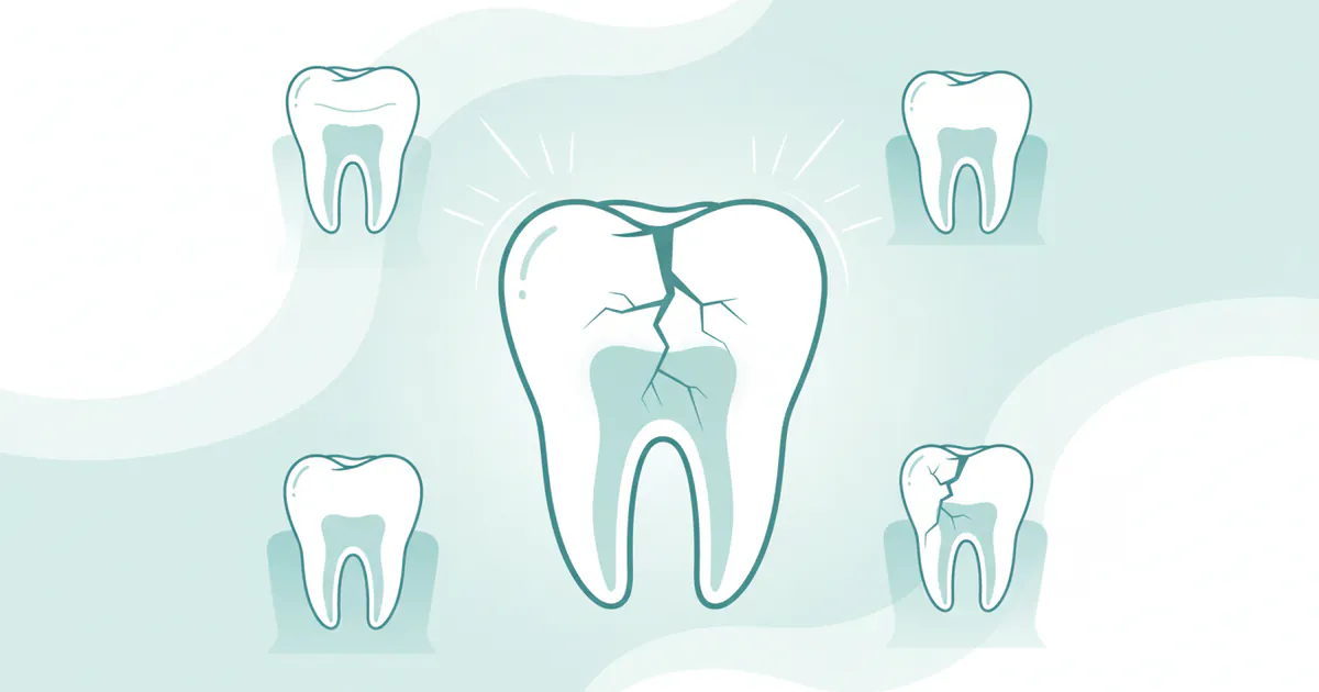
Progression to Larger Cavities:
- The most immediate risk is that the early demineralization will continue, breaking through the enamel to form a larger, visible hole (cavitation). Once this happens, remineralization alone is no longer an option, and a filling is required.
-
Dentin Involvement and Increased Sensitivity/Pain:
- As the cavity progresses deeper into the dentin (the layer beneath enamel), the sensitive tubules within the dentin become exposed. This leads to increased sensitivity to hot, cold, and sweet foods, and eventually, persistent tooth pain.
-
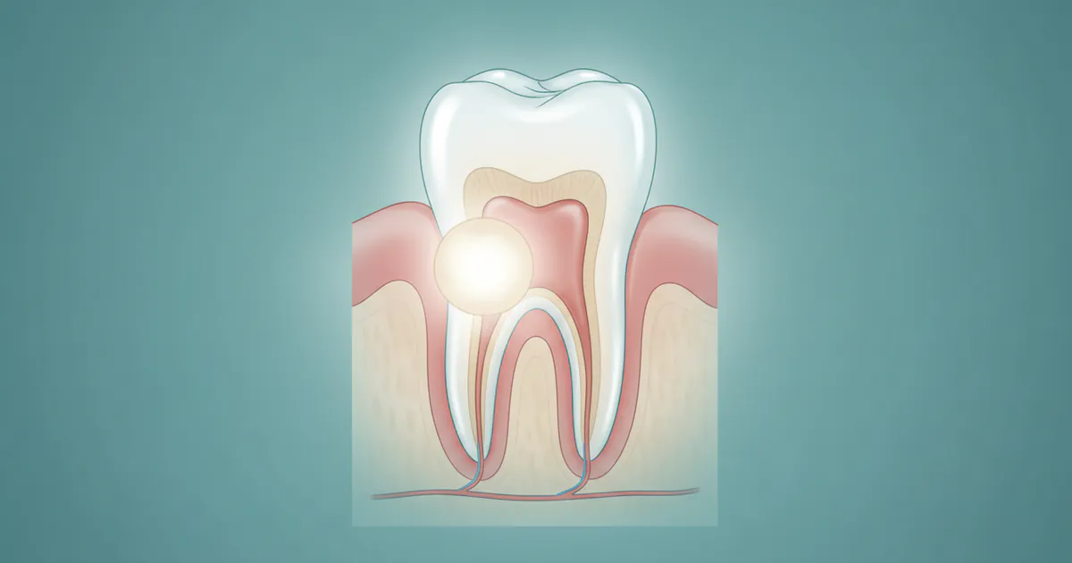
Pulpitis (Inflammation of the Nerve):
- If the decay reaches the pulp (the innermost part of the tooth containing nerves and blood vessels), it causes inflammation called pulpitis. This results in severe, throbbing tooth pain that can be constant and interfere with sleep.
- Reversible Pulpitis: Mild inflammation where the pulp can still heal if the decay is removed.
- Irreversible Pulpitis: Severe, often spontaneous pain where the pulp tissue is permanently damaged and dies. This typically requires a root canal treatment to save the tooth.
-
Dental Abscess (Tooth Infection):
- When the pulp dies, bacteria can multiply within the tooth, leading to an infection at the root tip or in the surrounding bone. This is a dental abscess.
- Symptoms: Severe, persistent pain; swelling in the face or jaw; fever; pus discharge; a "pimple" on the gums.
- Treatment: Requires immediate attention, often drainage, antibiotics, and either a root canal or tooth extraction.
-
Tooth Loss:
- If decay or infection is too extensive, the tooth may become unrestorable and need to be extracted. Missing teeth can lead to chewing difficulties, shifting of adjacent teeth, and bone loss in the jaw.
-
Systemic Infection (Rare but Serious): How Long Until a Tooth Infection Kills You?
- While extremely rare, an untreated dental abscess can spread beyond the jaw. Bacteria from a severe tooth infection can enter the bloodstream and travel to other parts of the body.
- Potential Complications:
- Cellulitis: A rapidly spreading bacterial infection of the soft tissues (face, neck).
- Ludwig's Angina: A severe form of cellulitis affecting the floor of the mouth and neck, which can obstruct the airway and become life-threatening.
- Cavernous Sinus Thrombosis: A blood clot in the cavernous sinus (a large vein at the base of the brain), which can cause vision loss, paralysis, and death.
- Sepsis: A life-threatening condition caused by the body's overwhelming response to an infection, leading to organ damage.
- Timeline to Fatality: There is no fixed "how long until a tooth infection kills you" timeline, as it depends on the individual's immune system, the virulence of the bacteria, and the specific site of spread. However, once a systemic infection like Ludwig's Angina or Sepsis develops, it can progress very rapidly, potentially becoming fatal within hours to days without aggressive medical intervention.
- The key takeaway is that while rare, a severe, untreated tooth infection is a medical emergency that can be fatal. It is never "just a toothache" when swelling, fever, or difficulty swallowing/breathing are present.
-
Impact on Overall Health:
- Chronic oral infections have been linked to systemic health issues, including heart disease, stroke, diabetes complications, and adverse pregnancy outcomes.
Therefore, addressing an early cavity is not just about saving a tooth; it's about safeguarding your overall health and well-being.
``
Children / Pediatric Considerations
Children are particularly susceptible to early cavities due to several factors, and addressing them is paramount for their developing oral health.

Why Children Are Susceptible:
- Thinner Enamel: Primary (baby) teeth have thinner enamel than adult teeth, making them more vulnerable to acid attacks and faster decay progression.
- Dietary Habits: Children often consume more sugary snacks and drinks.
- Developing Oral Hygiene: Young children may not have the dexterity or understanding to brush and floss effectively, requiring parental assistance.
- Deep Pits and Fissures: Their molars and premolars have naturally deep grooves where food and bacteria can easily accumulate.
Specific Considerations and Prevention for Children:
- Early Dental Visits: The American Academy of Pediatric Dentistry (AAPD) recommends a child's first dental visit by their first birthday or when their first tooth erupts. This helps establish a "dental home" and allows the dentist to assess risk and provide early guidance.
- "Lift the Lip" Exam: Parents should regularly lift their child's lip to check for white or brown spots along the gum line, especially on the upper front teeth, which can indicate early demineralization or "nursing bottle caries."
- Fluoride Varnish: Pediatric dentists frequently apply fluoride varnish during routine check-ups, often every 3-6 months, to strengthen young enamel.
- Dental Sealants: These are highly recommended for permanent molars as soon as they erupt (typically around ages 6 and 12) to protect the chewing surfaces from decay.
- Parental Assistance with Brushing: Parents should brush their child's teeth twice daily until they develop sufficient dexterity (usually around age 7-8). Use a rice-grain sized smear of fluoride toothpaste for children under 3 and a pea-sized amount for children 3-6.
- Dietary Guidance: Limit sugary drinks (including juice) and sticky snacks. Avoid putting babies to bed with bottles containing anything other than water. Encourage water consumption throughout the day.
- Silver Diamine Fluoride (SDF): SDF is an excellent non-invasive option for arresting early decay in primary teeth, especially for very young or uncooperative children who might not tolerate a traditional filling procedure. While it stains the decayed area black, it buys time and prevents further progression until the child is older or the tooth naturally exfoliates.
- Monitoring Primary Teeth: Even though baby teeth eventually fall out, they are crucial for speech development, chewing, and holding space for permanent teeth. Losing a primary tooth prematurely due to decay can lead to spacing issues and orthodontic problems later.
Pro Tip for Parents: Make brushing a fun, family activity. Use an age-appropriate brush and fluoride toothpaste. Consistency and leading by example are key to instilling good oral hygiene habits from an early age.
Frequently Asked Questions
What does an early cavity look like?
An early cavity, also called incipient caries, typically appears as a chalky white spot or a faint yellowish to light brown discoloration on the tooth surface. It indicates demineralization of the enamel, but usually, a visible hole (cavitation) has not yet formed. These spots may feel slightly rough if you run your tongue over them.
Can an early cavity heal itself?
Yes, in its very earliest demineralization stage, an early cavity can "heal" or remineralize. This process involves your saliva naturally depositing minerals back into the enamel, especially if aided by fluoride from toothpaste, water, or professional treatments. However, once a visible hole or cavitation has formed, the tooth structure is lost and cannot grow back, requiring a dental restoration like a filling.
Is an early cavity painful?
Generally, an early cavity is not painful. Pain usually indicates that the decay has progressed deeper into the dentin layer of the tooth, closer to the nerve. This is why regular dental check-ups are crucial, as a dentist can spot and treat early cavities long before you experience any discomfort.
How long does it take for an early cavity to become a big cavity?
The progression rate of an early cavity varies greatly depending on individual factors like diet, oral hygiene, saliva flow, and fluoride exposure. It can take anywhere from 6 months to several years for an early lesion to develop into a larger, cavitated cavity. Some early lesions may even remain stable for a long time or remineralize.
How much does it cost to fix an early cavity?
The cost depends on whether it's a non-cavitated lesion (treated with remineralization) or a small cavitated lesion (treated with a filling). Professional fluoride varnish costs $25-$75, and a small composite filling typically ranges from $100-$300 without insurance. With dental insurance, costs can be significantly reduced, often with fluoride treatments fully covered and fillings 50-80% covered.

Can I treat an early cavity at home?
You cannot treat an existing early cavity at home, but you can significantly aid in remineralization and prevent further progression. This involves rigorous at-home oral hygiene (brushing with fluoride toothpaste, flossing), using over-the-counter fluoride rinses, and improving your diet. However, a dentist needs to diagnose and recommend the appropriate professional treatment.
Is Silver Diamine Fluoride (SDF) a good option for early cavities?
SDF is an excellent non-invasive option for arresting the progression of early cavities, especially for very young children, individuals with special needs, or hard-to-reach areas. It effectively stops decay and promotes remineralization. The main drawback is that it permanently stains the decayed portion of the tooth black, which may be an esthetic concern for visible teeth.
What happens if an early cavity is left untreated?
If left untreated, an early cavity will continue to progress, leading to a larger cavity, increasing pain and sensitivity, potential infection of the tooth's pulp (nerve), and eventually a dental abscess. In severe, rare cases, a dental infection can spread to other parts of the body and become life-threatening.
Can I just wait until it hurts before seeing a dentist?
Waiting until a cavity hurts is a risky approach. Pain usually signifies that the decay has reached an advanced stage, potentially requiring more extensive, expensive, and invasive treatments like root canals or extractions. Early cavities can be treated minimally, often without drilling, and at a much lower cost. Prompt action saves your tooth, your wallet, and your overall health.
What foods cause early cavities?
Foods and drinks high in sugar and refined carbohydrates are the primary culprits. This includes soda, fruit juice, sports drinks, candy, pastries, cookies, and even starchy foods like bread and chips. Frequent snacking on these items, especially without brushing, creates a constant acidic environment that promotes early cavity formation.
When to See a Dentist: Don't Delay Care
Recognizing when to seek professional dental care is crucial for managing early cavities and preventing their progression. Here’s a clear guide:
Routine Care Guidance (Even Without Symptoms):
- Every Six Months: Schedule and attend routine dental check-ups and cleanings every six months. This is the most important step for catching early cavities. Dentists can detect demineralization or small cavitations visually or with X-rays long before you feel any symptoms.
- New Oral Hygiene Products: If you're considering new fluoride rinses, specialized toothpastes, or other preventive tools, discuss them with your dentist first.
Warning Signs That Need Prompt Attention (Not Necessarily Emergency):
- Visible White, Yellow, or Brown Spots: If you notice any unusual discoloration or chalky spots on your teeth that don't brush away, especially near the gum line or between teeth, schedule an appointment soon. These are classic signs of an early cavity.
- Persistent Mild Sensitivity: If you experience new or lingering mild sensitivity to hot, cold, or sweet foods/drinks, even if it's not severe pain, it could indicate demineralization or a small cavity reaching the dentin.
- Rough Spots on Teeth: If your tongue consistently catches on a rough spot on a tooth surface, it might be an area of enamel breakdown.
- "Catch" During Flossing: If your floss consistently snags or tears in the same spot between teeth, it could indicate a rough area or a developing cavity.
- Old Fillings/Restorations: If you notice a crack, chip, or discoloration around an existing filling, it could be a sign of recurrent decay underneath, warranting an evaluation.
Red Flags for Immediate Attention (Emergency/Urgent Care):
- Severe, Constant Tooth Pain: Especially if it's throbbing, prevents sleep, or doesn't respond to over-the-counter pain relievers. This indicates potential pulpitis or an abscess.
- Swelling in the Face, Gums, or Jaw: This is a strong indicator of a spreading infection or abscess and requires immediate dental or medical attention.
- Fever Associated with Tooth Pain/Swelling: A fever coupled with dental symptoms means the infection is spreading systemically.
- Difficulty Swallowing or Breathing: These are critical signs of a severe, potentially life-threatening infection (like Ludwig's Angina) and warrant an immediate trip to the emergency room or urgent dental care.
- Pus or a Pimple-like Bump on the Gums: This signifies an active infection draining from an abscess.
In summary, do not wait for pain to be your indicator. Early cavities are silent threats. Proactive, regular dental visits are your best defense against progression to more severe and costly problems. When in doubt, always consult your dentist.
Frequently Asked Questions
Medically Reviewed Content
This article was written by our dental health editorial team and reviewed for medical accuracy. Our content follows strict editorial guidelines for reliability and trustworthiness.
Medical Disclaimer
This article is for informational purposes only and does not constitute medical advice. Always consult with a qualified dental professional for diagnosis and treatment. Do not delay seeking professional advice because of something you read on this website.
Related Articles
What Is an Abscess
Imagine a throbbing pain deep within your jaw, a persistent ache that keeps you awake at night and makes even the softest foods unbearable. This isn't just a bad toothache; it could be a sign of something much more serious: a dental abscess. Affecting millions of Americans annually, an abscess is an
February 22, 2026
Cracked Tooth Syndrome: Complete Guide
Cracked Tooth Syndrome: Complete Guide Category: Dental Conditions & Diseases
February 22, 2026
Dentinogenesis Imperfecta: Complete Guide
Imagine a world where your teeth, fundamental to chewing, speaking, and even smiling confidently, are inherently fragile, discolored, and prone to rapid wear from the moment they emerge. For individuals affected by dentinogenesis imperfecta (DI), this is a reality. While relatively rare, affecti
February 22, 2026
Stage 1 Early Cavity: Complete Guide
Did you know that dental cavities are one of the most common chronic diseases globally, affecting both children and adults? In the United States, over 90% of adults have had a cavity, and a significant portion of these begin as a stage 1 early cavity. Often invisible and painless in its nasc
February 22, 2026