X-Ray (Dental)
5,620 words · 19 min read
Quick Definition
Diagnostic imaging that uses low-dose radiation to capture images of teeth, bones, and surrounding tissues, revealing problems not visible during a clinical examination.
X-Ray (Dental)
Short Definition: Diagnostic imaging that uses low-dose radiation to capture images of teeth, bones, and surrounding tissues, revealing problems not visible during a clinical examination.
Introduction
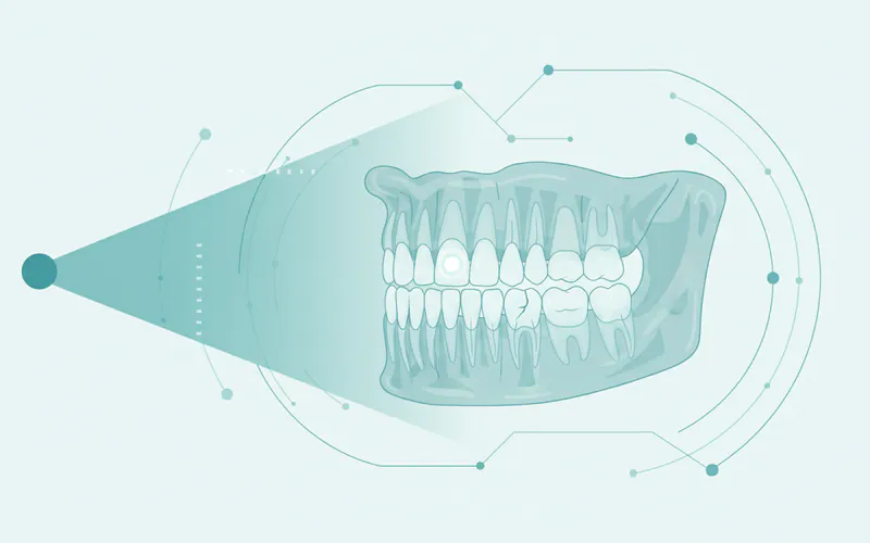
A dental X-ray, often simply referred to as an X-ray, is an indispensable diagnostic tool in modern dentistry. It employs a small, controlled dose of radiation to create detailed images of the inside of your mouth. Unlike what can be seen with the naked eye during a routine visual examination, dental X-rays penetrate beyond the surface, allowing dentists to visualize the internal structures of teeth, the bone supporting them, and the surrounding soft tissues. This capability is crucial for identifying a wide range of oral health issues, from hidden cavities and bone loss to impacted teeth and infections, long before they become symptomatic or visible.
The importance of dental X-rays for maintaining optimal oral health cannot be overstated. They are fundamental for early detection, enabling timely intervention that can prevent minor issues from escalating into major, painful, and costly problems. For instance, an X-ray can pinpoint a small cavity forming between teeth, allowing for a simple filling before it progresses to a deep infection requiring a root canal. It's also vital for planning complex procedures like placing dental implants or evaluating orthodontic needs.
Dental X-rays are a common and routine part of dental care for millions of Americans. According to the American Dental Association (ADA), the frequency of X-rays varies based on individual risk factors and dental history, but most adults will have bitewing X-rays taken every 1-3 years, while children and adolescents, due to their developing mouths and higher susceptibility to decay, may need them more often. This diagnostic practice significantly contributes to the overall health and longevity of your smile.
Key Takeaways:
- Early Detection is Key: Dental X-rays reveal hidden problems like cavities, infections, and bone loss not visible during a standard exam.
- Low Radiation Dose: Modern digital dental X-rays use minimal radiation, significantly less than in the past, making them very safe.
- Multiple Types for Different Needs: From bitewings for decay to panoramic for full jaw views and CBCT for 3D insights, specific X-ray types serve different diagnostic purposes.
- Foundation for Treatment: X-rays are crucial for planning treatments like fillings, root canals, and dental implants.
- Personalized Frequency: Your dentist determines how often you need X-rays based on your age, risk factors, and oral health history.
- Preventive Power: Regular X-rays help prevent minor issues from becoming major, painful, and expensive problems.
Detailed Explanation
Dental X-rays, often perceived simply as a snapshot, are in fact a sophisticated diagnostic tool with various applications, each designed to provide specific insights into your oral health. Understanding the different types, why they are recommended, and what to expect can demystify this essential part of your dental care.
Types and Classifications
Dental X-rays are broadly classified into two main categories: intraoral (taken inside the mouth) and extraoral (taken outside the mouth). Modern dentistry predominantly uses digital X-rays, which offer significant advantages over traditional film X-rays.
Intraoral X-rays: These are the most common type of dental X-rays and provide detailed images of individual teeth and the surrounding bone.
- Bitewing X-rays:
- What they show: Primarily used to detect decay between teeth (interproximal cavities) that are often impossible to see during a visual exam. They also show changes in bone level due to gum disease.
- How they're taken: You bite down on a wing-shaped device, holding the X-ray film or sensor in place. Typically 2-4 images are taken to capture all back teeth.
- Frequency: Often recommended annually or every 18-36 months for adults, and more frequently for children and individuals with a high risk of cavities.
- Periapical (PA) X-rays:
- What they show: Capture the entire tooth, from the crown (visible part) to the tip of the root and the surrounding bone. They are vital for diagnosing abscesses, bone loss around the root, root fractures, and issues related to root canal treatment.
- How they're taken: The sensor is placed vertically to capture one or two teeth.
- Frequency: Taken as needed, often when a specific tooth is causing pain or when evaluating the success of a prior treatment. A "full mouth series" (FMX) consists of 14-21 periapical and bitewing images to provide a comprehensive view of all teeth and surrounding structures, usually recommended every 3-5 years for new patients or as a baseline.
- Occlusal X-rays:
- What they show: Provide a broader view of an entire arch (upper or lower jaw), showing the full development and placement of teeth. Useful for detecting extra teeth, impacted teeth, jaw fractures, cleft palates, or large cysts and stones in the salivary glands.
- How they're taken: You bite down on a larger film or sensor.
- Frequency: Taken when necessary for specific diagnostic purposes.
Extraoral X-rays: These are taken from outside the mouth and provide a broader view of the jaw, skull, and surrounding structures.
- Panoramic X-rays (Panorex):
- What they show: Captures a single image of the entire mouth, including all teeth in both the upper and lower jaws, jawbones, temporomandibular joints (TMJ), and nasal and sinus areas. Excellent for detecting impacted wisdom teeth, evaluating jaw trauma, cysts, tumors, and planning for dentures or implants.
- How they're taken: The machine rotates around your head while you stand or sit still.
- Frequency: Typically recommended every 3-5 years or as needed for specific diagnostic purposes (e.g., orthodontic evaluation, wisdom tooth assessment).
- Cephalometric Projections:
- What they show: A lateral (side) view of the head, primarily used in orthodontics to assess the relationship of the teeth to the jaws and the jaws to the rest of the facial structure. Helps in treatment planning for braces.
- How they're taken: Taken with a specialized X-ray unit, similar to a panoramic machine.
- Cone Beam Computed Tomography (CBCT):
- What they show: Provides a 3D image of dental structures, soft tissues, nerve paths, and bone in a single scan. Invaluable for complex cases like precise implant placement, diagnosing TMJ disorders, evaluating nerve issues, planning extractions of impacted teeth, and assessing the extent of infections.
- How they're taken: A cone-shaped X-ray beam rotates around the patient, capturing hundreds of images that are then reconstructed into a 3D model.
- Frequency: Used selectively for complex diagnostic or treatment planning needs due to a slightly higher, though still low, radiation dose compared to other dental X-rays.
Digital vs. Film X-rays: Modern dental offices primarily use digital X-rays.
- Digital X-rays: Use an electronic sensor instead of film.
- Pros: Significantly lower radiation exposure (up to 80% less), instant image display, enhanced image quality (can be magnified and adjusted for contrast), easier storage and electronic sharing with specialists, and environmentally friendly (no chemical processing).
- Film X-rays: Traditional method using film that requires chemical processing.
- Cons: Higher radiation dose, slower processing time, chemical waste. While still effective, most practices have transitioned to digital.
Reasons Your Dentist Might Recommend an X-Ray (Formerly "Causes and Risk Factors")
Dental X-rays are not "caused" but rather are a necessary diagnostic step. The reasons a dentist recommends an X-ray are based on an assessment of a patient's individual needs, oral health status, and risk factors for dental problems.
Common Reasons for X-ray Recommendation:
- New Patient Comprehensive Exam: To establish a baseline of oral health, including a full mouth series (FMX) or panoramic X-ray to detect existing conditions, developmental issues, or hidden pathology.
- Routine Check-ups: Bitewing X-rays are often taken periodically to monitor for new cavities between teeth or changes in bone level indicative of gum disease.
- Signs of Decay or Infection: If you experience pain, sensitivity to hot/cold, visible stains or pits, or your dentist suspects decay based on a clinical exam, X-rays are crucial to confirm and determine the extent of the problem.
- Gum Disease Assessment: X-rays help evaluate bone loss around the teeth, a key indicator of periodontal (gum) disease.
- Orthodontic Evaluation: Panoramic and cephalometric X-rays are essential for planning braces or other orthodontic treatments, showing tooth position, jaw alignment, and growth patterns.
- Wisdom Teeth Assessment: Panoramic X-rays are used to monitor the development and eruption of wisdom teeth, identify impaction, or plan for extraction.
- Pre-surgical Planning: Before procedures like implant placement, tooth extractions, or root canal therapy, X-rays (especially CBCT scans for implants) provide detailed anatomical information to ensure safe and successful treatment.
- Evaluating Existing Restorations: To check the integrity of old fillings, crowns, or bridges, and to look for recurrent decay underneath them.
- Trauma or Injury: To diagnose fractures in teeth or jawbones following an accident or injury.
- Unexplained Symptoms: If you have persistent jaw pain, swelling, or other oral discomfort without an obvious cause, X-rays can help pinpoint the underlying issue.
Risk Factors for Needing X-rays More Frequently:
- High Cavity Risk: Individuals with a history of multiple cavities, poor oral hygiene, high sugar intake, or conditions causing dry mouth.
- Existing Restorations: Many fillings, crowns, or other dental work that require monitoring for recurrent decay or structural issues.
- Periodontal Disease: Patients with gum disease require X-rays to monitor bone loss progression.
- Developing Teeth: Children and teenagers with active tooth development, eruption issues, or high decay risk.
- Tobacco Use: Smokers and users of other tobacco products are at higher risk for gum disease and oral cancers, potentially requiring more frequent monitoring.
- Certain Medical Conditions: Diabetes, Sjogren's syndrome, or medications that cause dry mouth can increase the risk of dental problems.
Signs and Symptoms to Watch For (That Suggest the Need for a Dental X-Ray)
While X-rays are often taken proactively, certain signs and symptoms can indicate an underlying issue that warrants an immediate X-ray to accurately diagnose the problem. If you experience any of the following, it's a strong signal to consult your dentist:
- Persistent Toothache: Any lingering pain, whether sharp, dull, or throbbing, indicates a potential problem like a deep cavity, cracked tooth, or infection requiring an X-ray to locate the source.
- Sensitivity to Hot, Cold, or Sweet: While common, persistent or severe sensitivity can point to a worn enamel, exposed root, or a developing cavity that an X-ray can confirm.
- Visible Holes or Stains: Obvious dark spots or holes on your teeth are clear indicators of decay, but an X-ray will show how deep the cavity is and if it has reached the pulp (nerve).
- Swollen, Red, or Bleeding Gums: These are classic signs of gum disease, and X-rays are essential to assess the extent of bone loss associated with the condition.
- Loose or Shifting Teeth: Can be a sign of advanced gum disease or underlying bone issues, which X-rays can help diagnose.
- Bad Breath or Bad Taste: Persistent halitosis or a foul taste in your mouth, especially if accompanied by swelling, could indicate an abscess or infection that needs to be visualized with an X-ray.
- Swelling in the Jaw or Face: A serious symptom that could indicate a severe infection (abscess) or cyst, requiring immediate X-ray imaging for diagnosis and treatment planning.
- Difficulty Chewing or Biting: Pain when biting down can indicate a cracked tooth, deep cavity, or an issue with a filling or crown, all of which benefit from X-ray examination.
- Jaw Pain or Clicking: While TMJ disorders can have multiple causes, X-rays, including panoramic or CBCT, can help rule out underlying bone or joint pathology.
- Injury to the Mouth or Face: Following trauma, X-rays are critical to check for fractures in teeth, roots, or jawbones.
Diagnosis Process — What Your Dentist Does
When you visit your dentist for a check-up or specific concerns, the diagnostic process involving X-rays typically follows these steps:
- Clinical Examination: The dentist will first conduct a thorough visual and tactile examination of your mouth, teeth, and gums. They'll look for visible signs of decay, gum inflammation, existing restorations, and any other abnormalities. They will also ask about your medical history, symptoms, and concerns.
- Determine X-ray Needs: Based on the clinical exam, your dental history, risk factors, and any symptoms you're experiencing, the dentist will decide which type and how many X-rays are necessary. They follow guidelines from professional organizations like the ADA regarding frequency and necessity to minimize radiation exposure while maximizing diagnostic benefit.
- Preparation: You will be given a lead apron to wear, often with a thyroid collar, to protect your body from scattered radiation. This is a crucial safety measure, even with the low doses used in modern digital X-rays.
- Taking the Images:
- Intraoral X-rays: A small digital sensor (or film) connected to a computer will be placed inside your mouth. You may be asked to bite down on a small tab to hold it in position. The X-ray machine will be positioned next to your cheek, and the technician will step away briefly to activate the machine. This process is repeated for each image.
- Extraoral X-rays: For panoramic or cephalometric X-rays, you will stand or sit in a specific position, often with your chin resting on a support. The machine will then rotate around your head to capture the image. For CBCT scans, you'll typically sit or lie still while the scanner rotates.
- Image Review and Discussion: With digital X-rays, the images appear almost instantly on a computer screen. Your dentist will review these images, often magnifying them to show you any areas of concern. They will then explain their findings in clear, understandable language, pointing out issues like cavities, bone loss, cysts, or impacted teeth.
- Treatment Planning: Based on the X-ray findings and the clinical examination, your dentist will develop a comprehensive treatment plan, discussing the recommended procedures, their benefits, risks, and costs.
Treatment Options with Pros, Cons, and Costs
Dental X-rays diagnose problems; they do not treat them. The "treatment options" described here are for the conditions that dental X-rays commonly reveal. We will specifically address treatments related to the "related terms": Cavity, Root Canal, and Implant, along with other frequent findings.
1. Cavity (Dental Caries)
- Diagnosis by X-ray: Bitewing X-rays are excellent for detecting interproximal (between teeth) cavities, and periapical X-rays show the depth and extent of larger cavities.
- Treatment: Dental Fillings
- Description: The decayed part of the tooth is removed, and the remaining space is filled with a restorative material.
- Pros: Restores tooth function and aesthetics, prevents further decay, relatively quick procedure.
- Cons: Not permanent, may need replacement over time. Tooth sensitivity after filling is common but usually temporary.
- Types & Costs (US):
- Amalgam (Silver) Fillings:
- Pros: Durable, cost-effective.
- Cons: Silver appearance, mercury content (though considered safe by ADA), not bonded to tooth.
- Cost: $50 - $200 per filling.
- Composite (Tooth-Colored) Fillings:
- Pros: Aesthetically pleasing, bonded to tooth, less tooth structure removed.
- Cons: More expensive, may not be as durable for large fillings in high-stress areas, can stain over time.
- Cost: $100 - $350 per filling.
- Gold Fillings / Porcelain Inlays/Onlays:
- Pros: Very durable, aesthetically pleasing (porcelain).
- Cons: Most expensive, requires multiple appointments.
- Cost: $250 - $1,500+ per filling/inlay/onlay.
- Amalgam (Silver) Fillings:
2. Root Canal (Endodontic Treatment)
- Diagnosis by X-ray: Periapical X-rays reveal infections at the tip of the root (abscesses), internal decay, or inflammation in the pulp chamber.
- Treatment: Root Canal Therapy
- Description: When the pulp (nerve and blood vessels) inside a tooth becomes infected or inflamed, it's removed, the canal system is cleaned and disinfected, and then filled and sealed. A crown is almost always recommended afterward.
- Pros: Saves the natural tooth, prevents extraction, eliminates pain and infection.
- Cons: Can be expensive, requires multiple appointments (often 2), tooth may become brittle afterward and require a crown.
- Cost (US):
- Front Tooth (incisor/canine): $700 - $1,200.
- Premolar (bicuspid): $800 - $1,500.
- Molar: $1,000 - $2,000+.
- Note: These costs do not include the post and core or dental crown, which can add another $800 - $2,500.
3. Implant (Dental Implant)
- Diagnosis by X-ray: Panoramic X-rays show general bone volume, while CBCT scans provide critical 3D information on bone density, nerve pathways, and sinus locations, essential for precise implant placement planning.
- Treatment: Dental Implant Placement
- Description: A titanium screw is surgically placed into the jawbone to replace the root of a missing tooth. Once integrated with the bone, a crown, bridge, or denture can be attached.
- Pros: Most natural-looking and feeling tooth replacement, highly durable, preserves jawbone, does not affect adjacent teeth.
- Cons: High initial cost, surgical procedure, longer treatment time (healing period), not suitable for everyone (requires adequate bone).
- Cost (US):
- Single Implant (implant post only): $1,500 - $3,000.
- Abutment (connector): $300 - $600.
- Crown (on implant): $1,000 - $2,500.
- Total for single implant: $3,000 - $6,000+ (can be higher for complex cases, bone grafting).
Other Common Findings & Treatments:
- Gum Disease (Periodontitis): X-rays show bone loss around teeth. Treatment may involve scaling and root planing (deep cleaning), antibiotics, or gum surgery.
- Cost (scaling & root planing): $200 - $400 per quadrant.
- Abscess/Infection: Periapical X-rays show bone loss around the root tip. Treatment includes antibiotics, drainage, and often a root canal or extraction.
- Impacted Teeth (e.g., Wisdom Teeth): Panoramic X-rays reveal their position and relationship to other structures. Treatment is usually surgical extraction.
- Cost (wisdom tooth extraction): $75 - $200 (simple), $250 - $750 (surgical, impacted) per tooth.
- Cysts/Tumors: Various X-ray types, especially panoramic and CBCT, can detect these. Treatment ranges from monitoring to surgical removal.
Step-by-Step: What to Expect During Treatment (Illustrative Examples)
Since X-rays diagnose, "treatment" refers to what happens after the diagnosis. Here's a brief look at what you might expect for common procedures identified by X-rays:
1. For a Cavity (Dental Filling):
- Preparation: Local anesthetic is applied to numb the tooth and surrounding area. A dental dam may be placed to isolate the tooth.
- Removal of Decay: The dentist uses a high-speed handpiece (drill) to remove the decayed tooth structure.
- Preparation of Tooth: The cavity is shaped and cleaned to ensure the filling material adheres properly.
- Filling Placement: For composite fillings, a bonding agent is applied, followed by layers of resin material, which are hardened with a special light. For amalgam, the material is packed into the prepared cavity.
- Finishing: The filling is shaped, polished, and checked for proper bite (occlusion).
2. For a Deep Infection (Root Canal):
- Numbing and Isolation: Local anesthetic is administered. A rubber dam is placed around the tooth to keep it dry and free of saliva.
- Access Opening: A small opening is made through the crown of the tooth to access the pulp chamber.
- Cleaning and Shaping: Tiny instruments are used to remove the infected or inflamed pulp, and the root canals are cleaned and shaped.
- Disinfection: The canals are thoroughly irrigated with antimicrobial solutions.
- Filling the Canals: The cleaned canals are filled with a biocompatible material (gutta-percha) and sealed.
- Temporary Filling: A temporary filling is placed in the access opening.
- Crown Placement (Second Appointment): After healing, the tooth is prepared for a crown, which is essential to protect the now brittle tooth from fracture.
3. For a Missing Tooth (Dental Implant):
- Surgical Planning: Detailed CBCT scans are reviewed to determine the optimal implant size and position.
- Implant Placement Surgery: Under local anesthesia (and sometimes sedation), a small incision is made in the gum, a pilot hole is drilled into the jawbone, and the titanium implant post is carefully inserted. The gum is then sutured closed.
- Osseointegration (Healing): This is a critical period of 3-6 months where the implant fuses with the jawbone.
- Abutment Placement: Once integrated, a second minor surgery may be performed to attach a small connector post (abutment) to the implant. In some cases, the abutment is placed at the initial surgery.
- Crown Restoration: After the gum heals around the abutment, impressions are taken, and a custom-made crown is fabricated and cemented or screwed onto the abutment.
Recovery Timeline and Aftercare
Recovery and aftercare depend entirely on the treatment received as a result of the X-ray diagnosis.
- Dental Filling:
- Recovery: Numbness wears off in 1-3 hours. Mild sensitivity to hot/cold or pressure is common for a few days to a few weeks.
- Aftercare: Avoid chewing on the tooth until numbness subsides. Maintain good oral hygiene. Report persistent pain or sensitivity to your dentist.
- Root Canal Therapy:
- Recovery: Mild discomfort for a few days, managed with over-the-counter pain relievers. Swelling is rare but possible.
- Aftercare: Avoid chewing on the treated tooth until the permanent crown is placed to prevent fracture. Maintain excellent oral hygiene.
- Dental Implant Placement:
- Recovery: Soreness, swelling, and bruising for 3-7 days. Pain medication may be prescribed.
- Aftercare: Soft diet for several days. Avoid disturbing the surgical site. Good oral hygiene, carefully brushing around the implant site. Follow all post-operative instructions precisely, including antibiotic use if prescribed. Osseointegration (bone fusion) takes 3-6 months.
- General Aftercare for any Dental Procedure:
- Pain Management: Use over-the-counter pain relievers (ibuprofen, acetaminophen) as directed.
- Diet: Stick to soft foods initially, gradually reintroducing firmer foods. Avoid very hot, cold, or spicy foods.
- Oral Hygiene: Continue brushing and flossing gently, avoiding the surgical site if applicable. Your dentist may recommend a special mouth rinse.
- Follow-up Appointments: Attend all scheduled follow-up visits to monitor healing and complete treatment.
Prevention Strategies
The best way to minimize the need for extensive treatments (and the X-rays that diagnose them) is through proactive prevention.
- Maintain Excellent Oral Hygiene:
- Brush your teeth at least twice a day for two minutes each time with fluoride toothpaste.
- Floss daily to remove plaque and food particles from between teeth and under the gumline.
- Use an antiseptic mouthwash to reduce bacteria.
- Regular Dental Check-ups and Cleanings:
- Visit your dentist for professional cleanings and examinations typically every six months. These visits allow for early detection of problems before they become severe.
- Regular X-rays (as recommended by your dentist) are a vital part of these check-ups, revealing hidden issues.
- Balanced Diet:
- Limit sugary foods and drinks, which fuel decay-causing bacteria.
- Consume a diet rich in fruits, vegetables, and whole grains.
- Drink plenty of water, especially fluoridated water.
- Avoid Tobacco Products: Smoking and chewing tobacco significantly increase the risk of gum disease, oral cancer, and other dental problems.
- Use Fluoride: Fluoride strengthens tooth enamel and makes it more resistant to decay. This can be through fluoridated water, toothpaste, or professional fluoride treatments.
- Protective Gear: If you play contact sports, wear a mouthguard to protect your teeth from injury. If you grind or clench your teeth (bruxism), a nightguard can prevent excessive wear and potential fractures.
Cost Ranges in the US (With/Without Insurance)
The cost of dental X-rays and subsequent treatments can vary significantly based on location, the dentist's fees, and the complexity of the case. The following are general ranges in the US:
Cost of Dental X-rays:
| X-Ray Type | Without Insurance (Self-Pay) | With Insurance (Patient Portion) |
|---|---|---|
| Bitewing (2-4 images) | $30 - $80 | $0 - $40 |
| Periapical (single image) | $20 - $50 | $0 - $25 |
| Full Mouth Series (FMX) | $100 - $250 | $20 - $100 |
| Panoramic (Panorex) | $75 - $150 | $15 - $75 |
| CBCT (Cone Beam CT) | $300 - $700+ | $100 - $400+ |
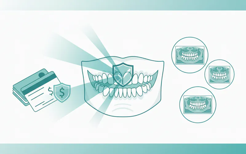
Impact of Dental Insurance: Most dental insurance plans cover preventive and diagnostic services, including dental X-rays, at a high percentage (often 80-100%). However, coverage limits, deductibles, and co-pays apply. For restorative treatments (fillings, root canals, crowns), coverage typically ranges from 50-80%, while major procedures like dental implants may have lower coverage or be excluded entirely. It's crucial to check with your specific insurance provider for exact details of your plan. Many dentists also offer payment plans or in-house discount programs for uninsured patients.
Comparison Table: Dental Filling Materials
| Feature | Amalgam (Silver) Fillings | Composite (Tooth-Colored) Fillings | Gold Fillings |
|---|---|---|---|
| Material | Mixture of metals (silver, mercury, tin, copper) | Resin and glass particles | Gold alloy |
| Appearance | Silver/dark gray, noticeable | Tooth-colored, blends with natural teeth | Gold, very noticeable |
| Durability | Very durable, lasts 10-15+ years | Good durability, lasts 5-10+ years (less for large fillings) | Excellent durability, lasts 15-20+ years |
| Cost (US) | $50 - $200 per filling | $100 - $350 per filling | $250 - $1,500+ per filling/inlay |
| Placement | Packs into cavity, requires minimal tooth removal | Bonded to tooth, conservative tooth removal | Lab-made, requires 2 appointments |
| Aesthetics | Poor | Excellent | Poor (for most) |
| Pros | Cost-effective, strong, long-lasting | Aesthetic, bonds to tooth, preserves tooth structure | Highly durable, biocompatible |
| Cons | Not aesthetic, mercury content (safe per ADA), can expand/contract | Less durable for large fillings, can stain, more expensive | Very expensive, noticeable, requires multiple visits |
| Best Used For | Back teeth where strength is paramount | Front teeth and smaller cavities on back teeth | Large restorations, high biting forces |
For Parents / Pediatric Considerations
Dental X-rays play a unique and crucial role in pediatric dentistry. Children's mouths are constantly changing, and X-rays help monitor growth and development, identify issues specific to primary and permanent teeth, and detect decay early in often hard-to-see areas.
- When are X-rays Needed for Children?
- New Patient: A set of bitewings or a panoramic X-ray may be taken to establish a baseline.
- Monitoring Growth & Development: To check for erupting permanent teeth, missing teeth, extra teeth, or jaw alignment issues. Panoramic X-rays are excellent for this.
- Cavity Detection: Children are often more susceptible to cavities, especially between teeth, due to diet and developing oral hygiene habits. Bitewing X-rays can detect these small cavities before they become large problems requiring fillings or even root canals on baby teeth.
- Trauma: To check for hidden tooth fractures or jaw injuries after an accident.
- Orthodontic Assessment: Critical for planning braces or other treatments for jaw and tooth alignment.
- Safety Concerns for Children:
- Parents often worry about radiation exposure for children. However, modern digital X-rays use extremely low doses. The benefits of early detection far outweigh the minimal risks.
- Dentists use lead aprons and thyroid collars to shield the child's body.
- The frequency of X-rays is tailored to each child's individual risk factors, dental history, and development stage, ensuring they are only taken when diagnostically necessary. The ADA recommends bitewing X-rays for children at high risk of decay every 6-12 months, and for children with low risk, every 12-24 months.
- Making it a Positive Experience: Dental offices are equipped to make X-rays quick and comfortable for children. Clear explanations, positive reinforcement, and a patient approach help children feel at ease.
Frequently Asked Questions
How much do dental X-rays cost?
The cost of dental X-rays in the US can range from $20-$80 for a single bitewing or periapical image, to $100-$250 for a full mouth series, and $75-$150 for a panoramic X-ray. A 3D CBCT scan is typically $300-$700+. These are self-pay prices; insurance usually covers a significant portion or all of these costs.
Are dental X-rays painful?
No, dental X-rays are not painful. The process involves placing a small sensor or film in your mouth, which might feel slightly uncomfortable or cause a brief gag reflex for some, but it does not cause pain. For extraoral X-rays like panoramic or CBCT, there is no discomfort as nothing is placed inside your mouth.
How long do dental X-rays take?
Taking a set of bitewing X-rays usually takes only a few minutes (5-10 minutes). A full mouth series might take 15-20 minutes. Panoramic X-rays are very quick, often less than 1 minute for the image capture itself. CBCT scans typically take 20-60 seconds. The entire appointment duration will depend on whether it's part of a larger examination.
Are there alternatives to dental X-rays?
While advanced technologies like intraoral cameras and visual examinations can help detect some issues, there is no direct alternative that can provide the same diagnostic information as a dental X-ray. X-rays are uniquely able to visualize hidden cavities between teeth, bone loss due to gum disease, infections below the gum line, impacted teeth, or problems within the jawbone, all of which are impossible to see visually.
Does dental insurance cover X-rays?
Yes, most dental insurance plans consider routine dental X-rays (bitewings, panoramic, full mouth series) as preventive or diagnostic services and typically cover them at a high percentage, often 80-100%, after any deductible is met. However, coverage limits (e.g., how often they're covered) and specific plan details vary, so it's always best to check with your insurance provider.
Are dental X-rays safe?
Yes, dental X-rays are considered very safe. Modern digital X-ray technology uses extremely low doses of radiation, significantly less than in previous decades. To further ensure safety, lead aprons and thyroid collars are used to shield the rest of your body from scattered radiation. The amount of radiation from a routine dental X-ray is comparable to the small amount of background radiation you're exposed to daily from the environment. The diagnostic benefits of early problem detection far outweigh the minimal risks.
How often do I need dental X-rays?
The frequency of dental X-rays is determined by your dentist based on your individual oral health needs, risk factors for disease, age, and symptoms.
- New patients: Often require a full mouth series or panoramic X-ray to establish a baseline.
- Adults with low risk: May need bitewing X-rays every 2-3 years.
- Adults with high risk (history of cavities, gum disease): May need bitewing X-rays every 6-18 months.
- Children/adolescents: May need bitewing X-rays every 6-12 months due to developing teeth and higher decay risk.
- Specific issues: Periapical, panoramic, or CBCT X-rays are taken as needed for specific diagnostic purposes, such as evaluating pain, planning implant placement, or assessing wisdom teeth.
Can I get a dental X-ray if I'm pregnant?
While the radiation dose from dental X-rays is very low, many dentists err on the side of caution and postpone routine X-rays until after pregnancy, especially during the first trimester. However, if a dental emergency or urgent diagnostic need arises (e.g., severe pain, infection), X-rays can be taken safely with appropriate lead shielding to protect both the mother and the baby. Always inform your dentist if you are pregnant or think you might be.
When to See a Dentist
Knowing when to seek dental care, whether routine or urgent, is key to maintaining your oral health and preventing more serious issues.
Routine Care (Schedule an Appointment):
- Regular Check-ups and Cleanings: The American Dental Association (ADA) recommends visiting your dentist at least once a year, and typically every six months, for a professional cleaning and examination. These appointments are crucial for preventive care and include necessary X-rays.
- New Patient Exam: If you haven't seen a dentist in a while or are new to an area, schedule a comprehensive new patient exam.
- Monitoring Existing Conditions: If you have ongoing gum disease, numerous fillings, or other chronic conditions, your dentist may recommend more frequent visits or specific X-ray intervals.
- Subtle Symptoms: Persistent bad breath, occasional sensitivity, or minor gum bleeding should prompt a visit to your dentist to investigate the underlying cause.
Urgent Care (See Your Dentist Soon):
- Persistent Toothache or Sensitivity: Any lasting pain, throbbing, or sensitivity to hot/cold/sweet that doesn't resolve can indicate a cavity, cracked tooth, or infection requiring an X-ray for diagnosis.
- Swollen Gums or Jaw: Swelling can be a sign of infection, gum disease, or an abscess.
- Bleeding Gums During Brushing/Flossing: While some light bleeding can occur, persistent or heavy bleeding indicates gum disease.
- Visible Holes or Dark Spots on Teeth: These are clear signs of decay that need immediate attention.
- Pain When Chewing or Biting: Could indicate a crack, deep decay, or issue with a restoration.
- Food Getting Trapped Between Teeth: Can signify a new cavity or a gap that needs addressing.
Emergency Care (Seek Immediate Dental Attention):
- Severe, Unbearable Tooth Pain: Excruciating pain, especially if accompanied by swelling, fever, or difficulty swallowing, indicates a serious infection or abscess.
- Facial Swelling: Rapidly spreading swelling in the face or jaw can be a sign of a life-threatening infection.
- Knocked-Out Tooth: Time is critical for saving a permanent tooth that has been knocked out. Seek care within 30-60 minutes.
- Broken or Chipped Tooth with Pain: If a significant portion of a tooth is broken, causing severe pain or exposing the nerve.
- Trauma to the Mouth or Jaw: Any injury resulting from a fall, accident, or blow to the face that causes pain, bleeding, or jaw misalignment.
- Uncontrolled Bleeding: Bleeding from the mouth that doesn't stop after applying pressure.
Prompt dental care, guided by the diagnostic power of dental X-rays, is essential for maintaining your oral health and ensuring a lifetime of healthy smiles.
Medical Disclaimer
This article is for informational purposes only and does not constitute medical advice. Always consult with a qualified dental professional for diagnosis and treatment. Do not delay seeking professional advice because of something you read on this website.